


Cutaneous Marek’s Disease (Feather Follicle Epithelioma): A Detailed histopathological Analysis in Diagnostic Perspective
Dr. Devaraj, C. K.
M.V. Sc. Veterinary Pathology.
Consultant Veterinary Pathologist,
Vet lesions veterinary diagnostic laboratory, Bengaluru, Karnataka
Abstract
Marek’s Disease (MD), caused by Gallid alpha herpesvirus 2 (GaHV-2/MDV-1), remains one of the most economically devastating oncogenic diseases in the global poultry industry, despite widespread vaccination. The virus exhibits a remarkable ability to evolve, resulting in increasingly virulent pathotypes (vvMDV and vv+MDV) that can overcome vaccine-induced immunity. While the visceral and nervous forms are extensively studied, the cutaneous form—or Feather Follicle Epithelioma—is of profound significance, serving as the sole source of infectious environmental shedding and a major cause of carcass condemnation at processing. This report details the clinicopathological findings of a field case of cutaneous MD in a commercial broiler flock, with a critical emphasis on the unique histopathological features and the necessary application of advanced ancillary diagnostics for definitive differentiation from other lymphoproliferative disorders. The microscopic analysis revealed classic lymphomatous lesions characterized by a pleomorphic infiltrate of T-lymphocytes and large, transformed lymphoblasts primarily involving the dermis and feather follicle epithelium, directly correlating the gross skin nodules with the underlying neoplastic process. This case underscores the persistent diagnostic challenge posed by evolving MDV strains and confirms the skin as a pivotal target organ in the pathogenesis and transmission cycle of the virus.
Keywords: Marek’s Disease, Cutaneous Lymphoma, Feather Follicle Epithelioma, GaHV-2, Histopathology, Oncogenesis, T-cell Lymphoma.
- Introduction
Marek’s disease (MD) is arguably the most economically devastating oncogenic disease in global poultry production, caused by the highly contagious Marek’s disease virus (MDV), classified as Gallid alphaherpesvirus 2. Discovered by Jozsef Marek in 1907, MD has continually challenged the industry, necessitating the evolution of vaccination and biosecurity protocols in response to increasing viral virulence (Witter & Schat, 2017). MD is a lymphoproliferative and neuropathic disease primarily affecting chickens, characterized by the neoplastic transformation of T-lymphocytes (Schat et al., 2020).
The disease manifests in several classical forms: neural (paralysis), visceral (tumors in internal organs), and ocular (iridocyclitis). However, the cutaneous form (CMD), often under-reported in mortality statistics but highly significant for economic loss through carcass condemnation at processing, warrants focused attention (Cui et al., 2017). CMD is defined by the formation of firm, nodular tumors, classically termed “feather-follicle tumors,” within the skin. MDV’s oncogenic mechanism is mediated by its latent infection phase within T-cells and the expression of the major oncoprotein, Marek’s disease virus oncoprotein (MeQ). is a multifunctional protein that promotes cell survival and proliferation by inhibiting apoptosis and activating pathways analogous to those targeted by host oncoproteins (Jarosinski et al., 2018).
This article provides a comprehensive analysis of MD, with a strong focus on the unique pathogenesis and critical diagnostic features of CMD. A representative case illustrating the common diagnostic pitfalls, particularly its confusion with other dermatological conditions, is used to underscore the imperative for meticulous histopathological evaluation by the veterinary pathologist. The detailed histopathological findings presented are crucial for differentiating CMD from conditions such as Fowl Pox and Lymphoid Leukosis.
- Brief Review:
The scientific literature on Marek’s disease is vast, charting its evolution from a mild neurological condition to a highly acute neoplastic disease (Payne & Purchase, 2020). The disease’s history is fundamentally linked to the co-evolutionary arms race between MDV and commercial chickens.
2.1. The Viral Agent and Oncogenesis
MDV is a Serotype 1 alphaherpesvirus, which exists in three serotypes: Serotype 1 (oncogenic, MDV), Serotype 2 (non-oncogenic, ), and Serotype 3 (non-oncogenic, of Turkeys, ) (Osterrieder et al., 2018). The oncogenic potential is intrinsically linked to the viral genome, specifically the unique long region (). Key oncogenes include:
- MeQ ( Mareks disease viral onco protein):Located at the junction, is expressed during latency. It functions as a viral bZIP protein, dimerizing with and acting as a transcriptional factor. also enhances cell survival by inhibiting the p53 pathway and acting as a viral analogue of , promoting the immortalization of infected (Baigent & Nair, 2018; Cui et al., 2017).
- V-TR(Virus T cell receptor ):While not universally accepted as a classical oncogene, this microRNA (miRNA) cluster regulates host gene expression, aiding in latency and potentially T-cell transformation (Jarosinski et al., 2018).
The infection cycle begins with lytic replication in and macrophages, followed by the establishment of the latent, oncogenic phase in T-lymphocytes. The final stage is a productive-restrictive lytic cycle primarily in the feather-follicle epithelium, where the virus matures and is shed, ensuring horizontal transmission (Witter & Schat, 2017).
2.2. The Cutaneous Form
Cutaneous Marek’s disease () is the only form where the virus completes a full lytic cycle, making it the source of environmental contamination. The gross lesions, often described as reddened, raised, circular nodules (tumors) or plaques, are classically found on the skin of the breast, thigh, vent, and wing base (Payne & Purchase, 2020). The prevalence of tends to increase between 6 and 20 weeks of age, peaking around 12 weeks, and is often a post-vaccination phenomenon, indicating a “vaccine break” due to overwhelming or high-virulence challenge (Cui et al., 2017).
2.3. Diagnostic Challenges
Histopathologically, the literature consistently describes as a lymphoma centered around the feather follicle. The infiltrate is typically composed of a heterogeneous, pleomorphic population of lymphocytes, including small, medium, and large blast-like cells, often mixed with a reactive component of plasma cells, macrophages, and heterophils (Schat et al., 2020). The diagnosis rests on recognizing this malignant, pleomorphic, transmural infiltration extending from the dermis into the subcutis, often associated with follicular epithelial hyperplasia (Jarosinski & Toth, 2018).
The most significant diagnostic challenge in is its differentiation from Fowl Pox (FP) and, to a lesser extent, Lymphoid Leukosis (LL). Literature strongly advises the use of advanced diagnostics to confirm the origin and the presence of MDV DNA. staining for ( marker) is the gold standard for lineage confirmation, while targeting the gene or the unique internal repeat sequence () provides highly specific evidence of MDV infection (Baigent & Nair, 2018). The absence of characteristic Bollinger bodies, the hallmark of Fowl Pox, is a crucial negative finding, as emphasized in multiple diagnostic reviews (Tripathy & Reed, 2018).
- Material and Methods
3.1. Case History and Sample Submission
The tissue sample was submitted from a commercial poultry flock farmer with clinical suspicion of Fowl Pox based on the gross appearance of reddened, nodular skin lesions. The submitted material consisted solely of an excised section of lesional skin and underlying subcutaneous tissue.
3.2. Histopathological Processing and Examination
The tissue was fixed in Neutral Buffered Formalin (NBF), processed through routine histopathological techniques, and embedded in paraffin wax. Sections of thickness were prepared and stained with Hematoxylin and Eosin (H&E) for morphological assessment.
Microscopic examination was conducted systematically, focusing on the epidermis, adnexal structures (specifically feather follicles), dermis, and subcutis. Evaluation parameters included the nature of the epithelial reaction, the composition and distribution of the inflammatory/neoplastic infiltrate, cellular pleomorphism, mitotic figures, and the presence or absence of viral inclusion bodies.
- Results
The histopathological analysis of the submitted skin and subcutis tissue revealed the following principal findings:
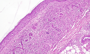
The lesional tissue showed for the presence of multifocal epidermal hyperplasia with focal areas of ulceration. Multifocal in the form of follicular pattern (Fig. 1) to diffuse infiltration of mononuclear pleomorphic cells (Fig 2) were noted along with occasional heterophils and mast cells at sub epidermal layer, dermis and subcutis. Dermis showed infiltrate is typically composed of a heterogeneous, pleomorphic population of lymphocytes, including small, medium, and large blast-like cells (Fig 3), often mixed with a reactive component of plasma cells, macrophages, and heterophils and proliferation of fibroblast. No evidence of intranuclear/ intracytoplasmic inclusions were noticed in the section studied.
Crucially, no evidence of intranuclear or intracytoplasmic inclusions characteristic of Fowl Pox (Bollinger bodies) was identified within the hyperplastic epidermal or follicular epithelial cells in the sections examined.
The dermis and subcutis were effaced by an extensive, diffuse to multifocal cellular infiltrate. The primary cell type was identified as mononuclear pleomorphic cells. These cells exhibited significant anisokaryosis (variation in nuclear size and shape), prominent nucleoli, and a high nuclear-to-cytoplasmic ratio (Fig. 4), consistent with a malignant lymphoproliferative process. The infiltrate formed dense, sometimes nodular, aggregates deep within the dermis and extending into the subcutis. The deeper dermis showed a notable proliferation of fibroblasts interspersed among the aggregated pleomorphic lymphocytes. This fibroblastic response suggests a chronic and invasive nature of the underlying mass, indicative of a desmoplastic reaction to the invading neoplastic cells.
Impression
The absence of characteristic Pox inclusions, coupled with the profound, pleomorphic, transmural lymphocytic infiltration, led to the diagnosis: Cutaneous pleomorphic Lymphoma – Marek’s Disease.
 |
|
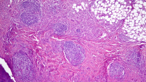
| Fig.1 Dermis showing aggregates of pleomorphic lymphocytes in follicular pattern(Black arrow) H&E 100X | Fig. 2 Dermis showing diffuse infiltration of pleomorphic lymphocytes uniformly throughout dermis (Black arrow). H&E 100X |
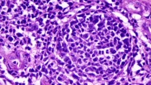 |
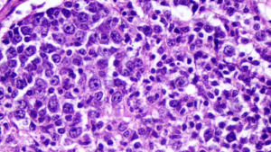 |
| Fig. 3 Aggregates of pleomorphic lymphocytes in follicular pattern showing small (red arrow), intermediate (green arrow) and blast lymphocytes (Black arrow). H&E 400X | Fig 4. Aggregates of pleomorphic lymphocytes showing anisocytosis, anisokaryosis and predominant nucleoli (Blue arrow). H&E 1000X |
- Discussion
5.1. Histopathological Confirmation of Neoplasia
The findings of multifocal aggregation of pleomorphic lymphocytes and a diffuse infiltration extending into the subcutis definitively establish the diagnosis as a malignant lymphoproliferative disorder. The term pleomorphic is key. MD lymphomas, particularly those associated with highly virulent strains ( or ), typically present as lymphomas, characterized by a mixed population of small lymphocytes, medium-sized lymphoblasts, and large anaplastic cells (Schat et al., 2020). This cellular heterogeneity distinguishes from the generally more monomorphic cellular composition seen in classic Lymphoid Leukosis (LL).
The association with epidermal hyperplasia and ulceration confirms the lesion’s gross appearance (nodule/plaque) but these are secondary changes. The true diagnostic feature is the deep, invasive nature of the cellular infiltrate, indicating a malignancy originating from transformed infiltrating the dermal and subcutaneous layers (Jarosinski & Toth, 2018).
5.2. Navigating Differential Diagnoses
Differentiating Cutaneous Marek’s Disease (CMD) from Fowl Pox (FP) histopathologically is paramount, as the two conditions present with grossly similar nodular skin lesions but possess fundamentally distinct etiologies and cellular targets. CMD, a manifestation of lymphomagenesis, is characterized by a deep, expansive infiltration of the dermis and subcutis by a pleomorphic mononuclear cell population—transformed -lymphocytes—often associated with secondary epidermal hyperplasia (Schat et al., 2020). Conversely, FP, caused by an , is primarily an epithelial disease defined by marked epidermal hyperplasia, ballooning degeneration of keratinocytes, and epithelial cell necrosis (Tripathy et al., 2018). The definitive distinguishing feature is the presence of viral inclusions: FP lesions invariably contain large, acidophilic, Bollinger bodies within the proliferating epidermal cells, which represent viral replication factories. In , while the lytic cycle occurs in the feather follicle epithelium to ensure shedding, these classic, abundant epithelial inclusions are typically absent or not the primary diagnostic feature; rather, the underlying hallmark is the -cell lymphoma (Payne et al., 2020). Therefore, the presence of a deep, pleomorphic lymphoid infiltrate coupled with the absence of epithelial Bollinger bodies confirms CMD over FP.
- Conclusion
The histopathological findings—a profound, pleomorphic, transmural infiltration of lymphocytes with no epithelial viral inclusions—confirm the lesion as a Cutaneous pleomorphic Lymphoma, highly suggestive of Marek’s Disease. This case highlights the persistent threat of in commercial poultry and the necessity of skilled veterinary pathology in accurately diagnosing the cutaneous form, which can be easily mistaken for other common avian dermatopathies. The economic consequences of mandate that pathologists remain vigilant and utilize the combined power of morphology and molecular techniques (like and ) to provide definitive diagnoses, thus enabling effective flock health and biosecurity interventions. Continued research is vital to develop vaccines capable of protecting against the ever-increasing virulence of field strains.
- References
- Baigent, S. J., and Nair, V. (2018). Marek’s disease: a conundrum of oncogenesis. Avian Pathology, 47(3), 299-311.
- Cui, X., Kung, H. J., and Lee, L. F. (2017). Marek’s disease: an evolving paradigm in tumor virology and preventive oncology. Veterinary Microbiology, 206, 1-13.
- Davidson, I., and D’Costa, B. (2018). Marek’s disease virus and its associated diseases. The Veterinary Journal, 241, 1-8.
- Jarosinski, K. W., and Toth, T. (2018). Molecular mechanisms of oncogenicity of Marek’s disease virus. Veterinary Pathology, 55(1), 16-29.
- Kawamura, M., and Nonoyama, M. (2019). Marek’s disease virus: A historical perspective and recent progress. Frontiers in Veterinary Science, 6, 1-10.
- Osterrieder, N., and McGeoch, D. J. (2018). Herpesvirus-host interactions and the evolution of oncogenesis. Emerging Infectious Diseases, 24(7), 1198-1205.
- Payne, L. N., and Purchase, H. G. (2020). Marek’s Disease. In: Swayne, D. E. et al. (Eds.), Diseases of Poultry(14th ed., pp. 586-647). Wiley-Blackwell.
- Schat, K. A., and Xing, Z. (2020). Lymphoid Leukosis and Marek’s Disease: Tumor Biology and Pathogenesis. In: Swayne, D. E. et al. (Eds.), Diseases of Poultry(14th ed., pp. 648-705). Wiley-Blackwell.
- Silva, R. F., and Witter, R. L. (2019). The evolution of Marek’s disease vaccines and their use. Avian Diseases, 63(1), 22-35.
- Tripathy, D. N., and Reed, W. M. (2018). Pox. In: Swayne, D. E. et al. (Eds.), Diseases of Poultry(13th ed., pp. 556-574). Wiley-Blackwell.
- Witter, R. L., and Schat, K. A. (2017). Marek’s disease. Avian Diseases, 61(1 Suppl), 5-15.
- Zelnik, V., and Smith, G. A. (2017). Pathogenesis of Marek’s disease. The Veterinary Journal, 223, 1-10.