


Field-based Diagnosis and Management of Canine Patellar Luxation in an Indian Veterinary Hospital
Palkhi Sharma (M.V.Sc, Veterinary Surgery & Radiology)
Introduction:
Hind limb lameness is one of the most common orthopaedic complaints encountered in small animal practice in India. Among the various causes, canine patellar luxation is frequently diagnosed, especially in toy and small breed dogs presented to veterinary hospitals. During my clinical training and postgraduate research at the Department of Veterinary Surgery and Radiology, College of Veterinary Science, Ludhiana, I had the opportunity to evaluate and surgically manage multiple cases of patellar luxation. This hands-on exposure offered valuable insight into real-life diagnostic challenges, clinical decision-making, client communication, and long-term management under practical hospital conditions.
The stifle joint plays a critical role in maintaining normal gait, balance, and limb stability. The patella functions as a mechanical lever that enhances the efficiency of the quadriceps muscle and enables smooth extension of the hind limb. When the patella fails to remain aligned within the femoral trochlear groove, it may displace either medially or laterally. This abnormal tracking alters joint biomechanics and can result in intermittent skipping gait, progressive lameness, pain, and, if left untreated, irreversible degenerative joint changes.
Patellar luxation is generally considered a developmental disorder. In routine clinical practice, small breeds such as Pomeranians, Chihuahuas, and Toy Poodles frequently present with medial patellar luxation, whereas larger breeds are more prone to lateral luxation. Clinical signs vary widely, ranging from mild occasional lameness to permanent disability depending on the grade of luxation and duration of the condition.
Over a period of 1 year, between March 2024 and February 2025, twelve dogs involving fourteen affected stifle joints were presented to our teaching hospital with complaints of hind limb weakness, abnormal gait, or recurrent lameness. These cases represented a spectrum of patellar luxation grades and provided an opportunity to observe diagnostic approaches, radiographic interpretation, surgical planning, rehabilitation strategies, and long-term outcomes in a real-world veterinary setting.
This article shares practical experiences gained from these cases, highlighting diagnostic strategies, field challenges, and clinical lessons that may help young veterinarians improve early recognition and management of patellar luxation while promoting animal welfare and responsible pet ownership.
Clinical Examination and Grading in Field Conditions:
Clinical examination was fundamental in aiding the diagnosis of dogs suspected of having patellar luxation. Most owners brought their pets with complaints such as intermittent skipping of a hind limb, reluctance to jump, difficulty climbing stairs, or gradually worsening lameness. Careful observation of gait at both walking and trotting speeds helped identify uneven weight bearing, altered stride length, and characteristic sudden flexion episodes that owners often described as “hopping” or “skipping.”
Palpation of the stifle joint in both standing and lateral recumbence positions allowed assessment of patellar stability, ease of displacement, joint smoothness, and pain response. In calm and cooperative dogs, gentle manual luxation and reduction of the patella helped determine the preliminary grade of the condition. However, in anxious, painful, or restless animals, accurate assessment was sometimes difficult without mild sedation, highlighting the importance of safe handling and patient comfort during orthopaedic examination.
Evaluating both hind limbs was essential, as several dogs showed early or subclinical involvement of the opposite limb. Muscle wasting, limb alignment, and subtle postural changes provided valuable clues regarding disease chronicity and biomechanical imbalance. In younger dogs, early deformities were often subtle and could easily be overlooked without thorough examination of the patient.
A key element in diagnosing patellar luxation is assigning an accurate grade, which required combining clinical findings with radiographic confirmation rather than relying on a single observation. In borderline cases, repeating the examination after analgesia or light sedation improved accuracy and confidence in decision-making. These experiences reinforced the value of a systematic and methodical orthopaedic examination in everyday practice.
Early and correct grading directly influenced treatment planning, prognosis discussions, and owner expectations. While mild cases could often be managed conservatively with monitoring and lifestyle advice, moderate to severe cases benefited from timely surgical correction to prevent long-term joint damage and chronic pain.
Radiographic Evaluation and Practical Observations:
Radiographic evaluation played a key role in confirming the diagnosis, understanding joint changes, and guiding surgical planning. In a busy teaching hospital setting, obtaining good-quality images often required patience, careful positioning, and clear coordination among clinicians, technicians, and handlers.
Each dog was assessed using three standard views of the stifle joint: cranio-caudal, medio-lateral, and skyline (Figure 1). Among these, the skyline view proved especially valuable for clearly visualizing patellar displacement and assessing the depth of the trochlear groove. In many cases, the patella could be clearly seen sitting outside its normal position, allowing confident differentiation between medial and lateral luxation. Shallow or flattened grooves were frequently observed, explaining the tendency for repeated displacement.
The cranio-caudal view helped evaluate limb alignment and detect early signs of degenerative joint changes such as osteophyte formation. Accurate positioning was critical, as even minor rotation could alter image interpretation. On several occasions, repeat radiographs were required to obtain diagnostic-quality views, reflecting a common practical challenge in routine clinical work.
The medio-lateral view was particularly useful for identifying joint effusion, assessing the patellar poles, and screening for concurrent orthopaedic problems. Mild to moderate osteophyte formation was observed in several cases, indicating ongoing joint stress. In a few dogs, erosive changes and subtle bone lesions were noted in advanced cases. One dog also showed features suggestive of cranial cruciate ligament involvement, reinforcing the need to always assess the joint comprehensively rather than focusing solely on the luxation.
Breed-related trends were also evident on imaging. Smaller breeds more commonly showed medial luxation with shallow grooves, whereas larger dogs frequently exhibited lateral luxation and femoral angulation.
Overall, radiography not only confirmed clinical suspicions but also supported surgical planning and improved communication with owners. These experiences highlighted the importance of careful positioning, thoughtful interpretation, and integrating imaging findings with physical examination for accurate diagnosis.

Figure 1: Cranio-caudal, skyline, and medio-lateral views of stifle joints showing lateral patellar luxation
Surgical Management and Intraoperative Experiences:
Surgical correction was recommended for dogs with persistent lameness, higher grades of luxation, or poor response to conservative management. Each case required individualized planning based on clinical severity, limb conformation, age, body weight, and owner expectations. Pre-operative discussions focused on explaining the procedure, recovery timeline, potential risks, and the importance of strict post-operative care.
All patients were prepared following standard aseptic protocols and underwent balanced anaesthesia with appropriate pain management. Intraoperative findings occasionally differed from preoperative imaging, reminding us that flexibility and sound anatomical understanding are essential during surgery.
The main surgical goals were to restore proper alignment of the quadriceps mechanism, deepen the trochlear groove when needed, and stabilize the patella within its natural tracking path. Depending on the case, a combination of techniques such as trochleoplasty, tibial tuberosity transposition, soft tissue release, and capsular tightening was performed. In younger dogs, special care was taken to protect growth plates and avoid excessive correction.
One of the recurring challenges was variability in bone quality and groove depth, especially in small breeds with long-standing luxation. Achieving adequate correction while preserving cartilage integrity required patience and precision. In advanced cases, adaptive bone changes and tight soft tissues made reduction more demanding and increased surgical time.
Accurate positioning of the tibial tuberosity was critical for long-term stability. Even minor deviations could affect post-operative comfort and patellar tracking. Testing joint movement during surgery by flexing and extending the limb helped confirm satisfactory alignment before final fixation.
In one case, suspected cranial cruciate ligament involvement required modification of the surgical plan and extended rehabilitation, highlighting the importance of complete joint assessment during surgery.
These surgeries strengthened clinical confidence, refined technical skills, and emphasized the value of careful planning and teamwork. Working in a teaching hospital also allowed meaningful interaction with interns and junior clinicians, fostering knowledge sharing and professional growth.
Post-Operative Care, Rehabilitation and Clinical Outcomes:
Post-operative care played a decisive role in determining the overall success of surgical correction for patellar luxation. All dogs received appropriate pain management, antibiotic coverage when indicated, and strict activity restriction during the early healing period. Owners were carefully guided on the importance of short leash walks, preventing jumping or climbing stairs, and maintaining proper wound hygiene. These simple but critical measures helped minimize complications and supported a smooth recovery.
Rehabilitation protocols were introduced gradually based on individual healing progress. Passive range of motion exercises, controlled weight-bearing, and progressive strengthening activities were demonstrated to owners to ensure correct execution at home. Compliance varied, particularly among first-time pet owners or those managing highly active dogs. Repeated counselling, visual demonstrations, and encouragement helped improve adherence and confidence in handling rehabilitation routines. In a few cases, home-based physiotherapy was supplemented with supervised sessions at the hospital to reinforce proper technique and motivation.
Most dogs showed visible improvement in limb usage within the first two weeks after surgery, with marked reduction in lameness by four to six weeks. Stable patellar tracking and improved gait symmetry were achieved in the majority of cases. Owners frequently reported increased activity levels, better comfort during movement, and overall improvement in their pets’ quality of life. Dogs that received early intervention generally recovered faster and demonstrated more consistent long-term functional improvement compared to those with chronic disease.
Minor post-operative issues such as localized swelling, temporary stiffness, and delayed return of full joint mobility were observed in a few dogs and were successfully managed with conservative care. One advanced case required prolonged rehabilitation due to concurrent joint pathology, reinforcing the importance of early diagnosis and timely surgical management to prevent secondary complications.
Regular follow-up visits allowed ongoing assessment of limb alignment, gait quality, and owner satisfaction. These interactions also created opportunities for reinforcing preventive care practices, educating owners about early orthopaedic warning signs, and promoting responsible breeding awareness in predisposed breeds. Over time, these follow-ups strengthened trust between clinicians and pet owners while encouraging long-term commitment to animal welfare.
Overall, these outcomes highlighted the value of structured rehabilitation, consistent follow-up, and active owner participation in achieving optimal recovery and sustained joint health.
Client Communication, Ethical Decision-Making, and Owner Education:
Effective communication with pet owners proved to be one of the most important components of successful case management. Many owners were unfamiliar with orthopaedic conditions and often underestimated the significance of intermittent lameness. Explaining the condition using simple language, diagrams, and radiographs helped bridge this knowledge gap and improved understanding and trust.
Discussing surgical costs, recovery expectations, and potential risks required sensitivity and transparency. Some owners expressed hesitation due to financial concerns or fear of anesthesia and surgery. Open dialogue regarding benefits, risks, and long-term welfare implications allowed owners to make informed decisions while ensuring ethical responsibility toward the animal. In selected mild cases, conservative monitoring was recommended when immediate surgery was not feasible and clinical signs were minimal.
Preventive education formed an important part of every consultation. Owners were encouraged to recognize early warning signs such as skipping gait, reluctance to exercise, and stiffness. Guidance on weight management, controlled exercise routines, and suitable home flooring helped reduce joint strain in predisposed breeds. These discussions often extended beyond individual cases and contributed to greater awareness within the pet-owning community.
Post-operative counselling required ongoing reinforcement. Written discharge instructions, live demonstrations of physiotherapy exercises, and scheduled follow-up visits significantly improved compliance and confidence. When owners actively participated in rehabilitation, outcomes were noticeably better, highlighting the importance of partnership between veterinarian and client.
These interactions strengthened interpersonal skills, patience, empathy, and adaptability in communication style. They also reinforced the understanding that clinical success depends not only on surgical skill but also on trust, education, and long-term collaboration.
Case Report: From Intermittent Lameness to Full Recovery (Figure 2):
A two-year-old male Pomeranian was presented to the hospital with a history of intermittent right hind limb skipping that had gradually worsened over six months. Initially, the owner assumed the behaviour was playful and harmless, delaying veterinary consultation until the dog began avoiding stairs and showing reduced activity levels. Clinical examination revealed that the patella could be manually displaced medially and remained luxated during flexion, indicating a moderate-grade patellar luxation.
Radiographic evaluation confirmed a shallow trochlear groove and medial displacement of the patella without advanced degenerative changes, making the case suitable for surgical correction. After detailed counselling regarding the procedure, expected recovery timeline, and post-operative responsibilities, the owner agreed to proceed with surgery.
Surgical correction involved deepening of the trochlear groove combined with soft tissue realignment. Intraoperatively, tightening of the medial joint capsule was observed, which required careful release to restore balanced patellar tracking. Stability was confirmed through repeated flexion and extension testing before closure.
Post-operatively, strict activity restriction, pain management, and gradual physiotherapy were implemented. Initial compliance was challenging due to the dog’s energetic temperament, requiring repeated counselling and regular follow-up communication. Over the following six weeks, steady improvement in weight bearing and gait symmetry was observed. By three months, the owner reported complete resolution of lameness and a return to normal daily activity.
This case highlighted the value of early intervention, effective owner education, and structured rehabilitation in achieving favourable outcomes. It also demonstrated how timely veterinary guidance can prevent long-term joint damage and improve quality of life for companion animals.

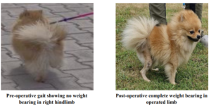
Figure 2: Pre- and post- operative images of surgically treated two year old Pomeranian
Field Challenges and Practical Lessons Learned:
Managing orthopaedic cases such as patellar luxation in an Indian veterinary hospital involves many practical challenges that extend far beyond the surgical procedure itself. One of the most frequently encountered issues was delayed presentation. Many pet owners initially interpreted intermittent skipping, mild limping, or altered posture as temporary or harmless behaviour. Veterinary attention was often sought only when the problem became persistent, painful, or visibly disabling. By this stage, secondary joint changes, muscle wasting, and adaptive bone remodelling had already developed, making both diagnosis and surgical correction more complex. These encounters highlighted the critical importance of early detection and proactive owner education.
Client awareness and compliance emerged as equally important factors influencing clinical outcomes. Explaining the anatomical nature of patellar luxation in simple terms, clarifying why surgery may be required, and emphasizing the importance of post-operative restriction demanded patience, clarity, and empathy. Some owners struggled to restrict highly active pets, particularly in limited living spaces or busy household environments. Others faced difficulties maintaining regular physiotherapy routines due to work commitments or lack of confidence in performing exercises correctly. Visual demonstrations, written instructions, repeated counselling, and regular follow-up calls proved valuable in improving understanding and adherence. These experiences reinforced the importance of communication skills as a core clinical competency.
Resource and infrastructure limitations represented another practical consideration in a teaching hospital setting. Advanced diagnostic imaging, specialized implants, and long-term rehabilitation facilities may not always be accessible or financially feasible for every client. This required careful prioritization and ethical decision-making to achieve the best possible outcomes using available resources. Thoughtful use of standard radiography, precise surgical technique, and realistic rehabilitation planning allowed effective care without compromising animal welfare. Balancing affordability with quality care became an essential aspect of responsible veterinary practice.
Patient-related challenges also influenced clinical management. Toy breeds often presented with fragile bone quality and narrow anatomical structures, requiring delicate handling and precise instrumentation during surgery. Chronic cases demonstrated adaptive changes in bones and soft tissues that increased technical difficulty and extended surgical time. Additionally, anxious or aggressive animals posed challenges during imaging, handling, and post-operative care. Managing such patients safely required teamwork, calm handling techniques, and coordinated efforts among clinicians, interns, and support staff.
From a professional perspective, each case strengthened the importance of clinical judgment, adaptability, empathy, and communication alongside technical competence. Learning to make balanced decisions under constraints, handling uncertainty with confidence, and supporting anxious owners became valuable life skills beyond surgical expertise. Over time, these experiences improved confidence in independent decision-making, sharpened problem-solving abilities, and deepened understanding of the emotional dimensions of veterinary care.
These experiences reinforced that successful field practice is not defined solely by surgical outcomes, but by holistic case management, ethical responsibility, client partnership, and collaborative teamwork. The ability to adapt to real-world limitations while maintaining high professional standards remains a defining strength of effective clinicians.
Reflections and Conclusion:
Working with canine patellar luxation cases in a real-world hospital environment proved to be a deeply transformative professional journey. Beyond strengthening surgical skills, these cases enhanced clinical reasoning, decision-making under pressure, communication abilities, and a greater appreciation for patient-centred care. Each successful recovery brought not only professional satisfaction but also reassurance that timely intervention can significantly improve quality of life for animals and peace of mind for their owners.
As a young woman veterinarian working in a physically demanding and technically intensive surgical discipline, these experiences fostered confidence, resilience, and professional identity. Orthopaedic surgery often requires stamina, precision, leadership, and mental focus. Navigating self-doubt, earning the trust of clients and colleagues, and learning to take responsibility for complex clinical decisions contributed greatly to personal growth. Participating in case discussions, mentoring junior trainees, and collaborating within multidisciplinary teams further strengthened professional maturity and leadership skills.
The lessons learned from these field-based experiences highlight the importance of early diagnosis, ethical decision-making, preventive education, and accessible veterinary services in improving long-term outcomes for companion animals. Increasing awareness among pet owners, encouraging timely veterinary consultation, promoting responsible breeding practices, and strengthening referral systems can significantly reduce the burden of preventable orthopaedic disease in India.
Inspired by the legacy of Savitribai Phule, whose courage and commitment to education challenged social barriers and empowered generations, women professionals today continue to reshape science, healthcare, and community development through dedication and perseverance. In veterinary medicine, women play an increasingly vital role in advancing animal welfare, supporting livelihoods, mentoring future professionals, and contributing to national development goals.
This journey reinforces that meaningful impact in veterinary practice is built not only on technical excellence, but also on empathy, integrity, leadership, and lifelong learning. By combining clinical competence with compassion and social responsibility, veterinarians can create lasting change for animals, communities, and the profession itself.