

SURGICAL MANAGEMENT OF A MID-SHAFT TIBIAL FRACTURE IN AN 8-YEAR-OLD INDIGENOUS FEMALE DOG: CASE REPORT
COLLEGE OF VETERINARY SCIENCE AND ANIMAL HUSBANDRY, NDVSU, JABALPUR, MADHYA PRADESH, INDIA
JAGMOHAN RAJPUT, ATUL S. PARIHAR, VIVEK K. MAURYA, ASHOK K. PATIL AND SHASHANK VISHVAKARMA
Corresponding Author – Jagmohan Rajput
Email- drjagmohanrajput07@gmail.com
ABSTRACT
This report presents the successful management of a mid-shaft tibial fracture in an 8-year-old indigenous female dog weighing 15.6 kg in a private clinic, Indore, (M.P). The dog exhibited non-weight-bearing lameness in the affected limb. Surgical stabilization was performed using a 3.5 mm intramedullary pin, following aseptic bone preparation with transverse flattening of the fracture ends. The procedure was conducted under balanced anaesthesia with Butorphanol, and Propofol, maintained with Isoflurane. Post-operative care included inj. Amoxicillin-Clavulanic acid, Meloxicam and regular antiseptic dressing. A fiber cast was applied to control axial rotation. The dog recovered fully within 40 days.
KEY WORDS: Butorphanol, Intramedullary, Propofol and Tibial Fracture.
INTRODUCTION
Intramedullary pinning is one of the oldest and acceptable methods of internal fracture fixation. The technique is popular as the implant is cost effective, user friendly and more biological but it resists only bending forces acting across the site of fracture; thus, simple intramedullary pinning is frequently associated with certain postoperative complications such as migration of pin from site of insertion, nerve damage, breakage of pin etc. Fractures of the long bones are commonly encountered orthopedic emergencies in veterinary practice, particularly in active and aging canine patients. Among these, tibial fractures represent a significant proportion due to the tibia’s subcutaneous location, especially along its medial surface, which renders it more susceptible to trauma (Johnson et al., 2005). Mid-shaft fractures of the tibia are frequently caused by vehicular accidents, falls, or blunt force trauma and can vary in complexity depending on the extent of soft tissue involvement and fracture pattern (Piermattei and Flo, 2006).
Surgical intervention is often warranted in mid-diaphyseal tibial fractures to ensure proper alignment, stability, and timely healing, especially in adult dogs where conservative management may not yield satisfactory functional outcomes (Millis & Levine, 2014). Internal fixation using techniques such as intramedullary pinning, plating, or external skeletal fixation is chosen based on the fracture type, patient’s size, and surgeon’s expertise. Tibial fractures, particularly diaphyseal (mid-shaft) ones, are among the most frequently encountered long-bone injuries in dogs, often resulting from high-energy trauma like traffic accidents (Dhongade et al., 2024). Among surgical options, intramedullary (IM) pinning remains a cornerstone technique due to its simplicity, cost-effectiveness, minimal soft-tissue disruption, and promotion of “biological” secondary bone healing.
Steinmann pins (with threaded tips) facilitate better rotational control and earlier weight-bearing compared to plain pins, with fewer complications such as pin migration and seroma formation (Dhongade et al., 2024). However, evidence remains limited regarding optimal fixation in skeletally immature dogs, and high-level studies comparing internal vs external methods. This underscores the continued relevance of IM pinning, especially where resources or advanced implants are limited.
CASE HISTORY AND CLINICAL PRESENTATION
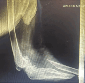
An 8-year-old indigenous female dog (Desi breed) weighing 15.6 kg was presented with an inability to bear weight on the right hind limb. Clinical and radiographic examination confirmed a mid-shaft tibial fracture. Due to the severity of the fracture, surgical stabilization was deemed necessary.
Anesthesia and Surgical Procedure
Anesthetic Protocol:
- Pre-medication: Butorphanol (0.2 mg/kg IV)
- Induction: Propofol (4 mg/kg IV)
- Maintenance: Isoflurane (1.5%) via inhalation anesthesia
Surgical Approach and Fracture Management:
- Aseptic Preparation and Incision: A standard aseptic technique was followed, and a medial incision was made to expose the fractured tibial ends.
- Bone Preparation: The fracture ends were transversely flattened, and bone marrow debris was removed to ensure proper pin insertion.
- Intramedullary Pinning: A 3.5 mm intramedullary pin was inserted using a retrograde approach, followed by normograde placement for enhanced stability.
- Wound Closure and Casting: The surgical site was closed using absorbable and non-absorbable sutures in a routine manner. A fiber cast was applied to immobilize the limb while keeping a window at the wound site for dressing. The cast was maintained for 12 days to prevent axial rotation and allow proper healing.
Post-Operative Management:
- Analgesia: Meloxicam (0.2 mg/kg SC)
- Antibiotics: Amoxicillin-Clavulanic acid (20 mg/kg IM, BID for 7 days)
- Gastroprotection: Pantoprazole (1 mg/kg PO)
- Wound Care: Regular antiseptic dressing through the cast window
- Monitoring: The dog exhibited progressive weight-bearing improvements, with full recovery observed within 40 days.

CONCLUSION
The combination of intramedullary pinning and external fiber casting provided effective stabilization, facilitating fracture healing. Proper aseptic preparation, flattening of fracture ends, and post-operative care played crucial roles in successful recovery. This case demonstrates the efficacy of surgical intervention in tibial fractures in indigenous dogs, emphasizing the importance of stabilization techniques and pain management for optimal outcomes. Recent comparative studies have emphasized the biomechanical and clinical benefits of using end-threaded Steinmann pins, which enhance rotational stability and reduce postoperative complications (Dhongade et al., 2024). While more advanced fixation options like locking plates or external skeletal fixators may be indicated in complex fractures, intramedullary pinning remains an effective modality for simple diaphyseal fractures, particularly in non-descript or medium-sized breeds.
This case reinforces the continued relevance of traditional orthopedic techniques like IM pinning, which, when applied correctly, can yield excellent clinical outcomes with minimal invasiveness.
REFERENCES
- Aydın, U., Özaydın, İ., Aksoy, Ö., Ermutlu, Ç. Ş., Kılıç, E., Yıldız, U. and Tanrıverdi, E. (2022). Clinical and radiological evaluation of tie-in osteosynthesis with intramedullary threaded pin in diaphyseal humeral, tibial, and femoral fractures in dogs. Kafkas University Veterinary Faculty Journal, 28(5), 593–599.
- Chitty, J., and Aldridge, P.(2024). Comparing internal versus external fixation for diaphyseal tibial and fibular fractures in skeletally immature dogs. Veterinary Evidence, 9(1).
- Dhongade, R. M., Bhadane, B. K., Akhare, S. B., Fiske, G. A. and Kamble, M. V. (2024). Comparative evaluation of plain and end threaded Steinmann pin for intramedullary pinning oflong bone fractures in dogs. Int. J. Vet. Sci. Anim. Husbandry, 9(4):517–21.
- Gupta, N. and Kumar, A. (2023).Elastic osteosynthesis in dogs – A Review. Pharma Innovation, 12(6):1021–1025.
- Johnson, K. A., Houlton, J. E. F. and Vannini, R. (2005). AO Principles of Fracture Management in the Dog and Cat. AO Publishing.
- Millis, D. L. and Levine, D. (2014). Canine Rehabilitation and Physical Therapy(2nd ed.). Elsevier Health Sciences.
- Piermattei, D. L. and Flo, G. L. (2006). Handbook of Small Animal Orthopedics and Fracture Repair(4th ed.). Saunders Elsevier.