


BOVINE ACTINOMYCOSIS/ LUMPY JAW DISEASE IN LIVESTOCK
Post no-1438 Dt 17/10/2019
by- Dr. Manoranjan Nanda, Veterinary Asst Surgeon, Bhubaneswar,
Dr. Jagadish Mohanty & Dr. Durga Pd. Das (Phd),Bhubaneswar, Odissa.
Introduction:
Actinomycosis, commonly called ‘Lumpy Jaw’ , usually affects the bony tissues of the head rather than the soft tissues (as opposed to Wooden Tongue). It is caused by infection with Actinomyces bovis, which is part of the normal bacterial flora of the upper digestive tract. The bacteria usually invade the skin through a wound or minor trauma caused by sticks, straw or barley awns, or when deciduous teeth are being shed.
Actinomycosis has been recorded from various parts of India. The incidence in cattle is higher where they are fed with straw and ensilage. These feeds injure the buccal mucosa and there by predispose them to infection.
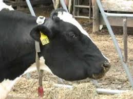
Actinomycosis is chronic, progressive, pyogranulomatous rarefying osteomyelitis of the maxillae, mandible, or any other bony tissues of head. The mass will be slow in growth, firm in consistency, painless and attached to the mandible. The alveoli of the roots of the cheek teeth are involved and results to loose teeth which makes chewing difficult and painful. Painful chewingresults to evidentweight loss.Ulceration occurs with or without tracts draining purulent material. These organisms are part of normal flora of the gastrointestinal tract of ruminants, they enter inside tissues following trauma to mucosa through abrasions and penetrating wounds may be from wire, sharp branches, coarse hay or sticks. Bacteria may also invade tissue through dental alveoli during tooth eruption. The defining feature of actinomycosis is the presence of a non-painful swelling under the jaw. This swelling can rupture and drain pus-type, smelly fluid which contaminates the environment.
Causative agent:
Actinomyces bovis is responsible for actinomycosis/lumpy jaw, which is a gram-positive, non-spore forming, non-motile, non-acid fast, facultative anaerobic pleomorphic coccobacillary bacterium in the genus Actinomyces.
Host:
Cattle are mainly affected. Pigs, sheep and goats are affected occasionally.
Zoonotic importance:
Actinomyces bovis is a zoonotic organism, causes granulomas, abscesses, skin lesions, and bronchopneumonia in humans.
Clinical signs:
• Initially Actinomyces bovis infection of the mandible or maxilla appear as warm, painful swellings consisting of distinct oedema overlapping a firm, painful, bony swelling which can easily be mistaken as traumatic injury.
• After a week or two, infection gets settle in bone, enlargement becomes more hard, painful and soft tissue oedemabecomes less apparent.
• Untreated cases results to pyogranulomatous infection of bone and associated soft tissues, which ultimately develop to granulomas.
• Draining tracts forms through the skin or into the oral cavity. These tracts discharge copious quantities of serous or mucopurulent pus that can be source of infection to other animals.
• Radiographs of skull confirm severe osteomyelitis with multifocal radiolucency caused by rarefaction of bone.
• Because of distortionof teeth, eating becomes more difficult for severely affected cows.
• Salivation, reduced appetite, hesitant attempts to chew, and dropping food from the mouth may be observed.
• Oral mucosal or tongue lacerations may be apparent.
• Hard, immobile, bony mass on the mandible
• Late in the disease, draining tracts may erupt
• Inflammation of the lymph nodes (lymphadenitis)
• Swelling of the pharynx (back of the throat)
• Excessive salivation due to pain
• Abnormal eating, dropping of feed from the mouth, inability to grasp food properly
• Fever in the early stages of disease
Diagnosis:
• Based on clinical signs, location of lesion and species involved
• Radiographs of head (determines degree of bone destruction)
• Isolation and identification of Actinomyces bovis in laboratory (specimen: exudates, aspirates, tissue samples). Actinomyces bovis shows MZN (modified Zeil-Neelsen) negative staining and non-haemolytic growth on media.
• Histopathological examination of specimens from lesions reveals aggregates of filamentous organisms surrounded by typical eosinophilic club-shaped structures
• Differential diagnosis should be done from actinobacillosis which also involve soft tissue of head (wooden tongue).
Control strategy & Prevention of Lumpy Jaw–
As the Actinomyces bovis is consisting normal flora of GIT and oropharynx in cattle, control of lumpy jaw focuses on avoiding coarse, poor quality stemmy feeds/hay or feeds with plant awns that might damage the mucosal U epithelium and allow entry of bacteria to soft tissue.
Rough grazing can be a risk factor for lumpy jaw as it may cause trauma, allowing the normal flora bacteria Actinomyces bovis to establish infection in the bony tissues of the mouth.
Avoiding rough grazing, soil-contaminated silage and “weedy” hay/haylage or straw for cattle feed usually prevents trauma to mucosal membranes in the mouth.
In an outbreak, it is important to identify the causative factors of the predisposing lacerations in the mouths of the affected animals. This is likely to be a feed source that, once identified, should be removed. Again, humane slaughter of affected animals is necessary at the appearance of clinical symptoms.
As the causative bacteria do not survive long in the environment and are present in the mouths of healthy cattle, the removal of the predisposing feed source is adequate for controlling the disease within a herd. If the problem persists, it is important to look for further sources of predisposing factors and eliminate them.
Treatment:
Purpose of the treatment is to kill bacteria and stop its transmission to other body tissue and animals.Duration of therapy is dependent on the severity of the lesion and response to therapy
• Sodium iodide (treatment of choice in ruminant) @ 70 mg/kg of a 10%–20% solution, IV) is given once and repeated several times at 7to 10day intervals. If any kind of Iodine toxicity (dandruff, diarrhoea, anorexia, flaky skin coughing, and excessive lacrimation) is observed, Iodine administration should be discontinued or given at long intervals
• Prolonged therapy with penicillin or penicillin-streptomycin (22,000 U/kg once daily), parenterallyfor at least 7 days
• Isoniazid per os for 30 days is also recommended
• When lesions are small and circumscribed, surgery is the treatment of choice
• Surgical debulking or removal of large pyogranulomas projecting from the skin of advanced cases may reduce the size of the lesion.
• Loose teeth may require extraction, and fistulous tracts may be flushed with iodine solution.
TREATMENT AT THE FIELD LEVEL———
Actinomyces bovis found sensitive to Penicillin, Streptomycin, Tetracycline, Bacitracin, Cloxacin and Co-trimoxazole. Dicrystin- DS is also sensitive . The affected animals are treated with injection penicillin along with Streptomycin Sulphate at the rate of 10 mg / kg body weight (Dicrystin-S,) daily along with Potassium Iodide 10 gram orally daily for 7 to 10 days. Local dressing with Povidone Iodine (Betadine, WineMedicare pvt. ltd.) of the wounds in the mandible region is done daily till the local healing of wounds is completed. Treatment of actinomycosis with Streptomycin combined with Potassium Iodide at the rate of 6-10 gm /day orally for 7-10 days is also been found effective. Oral administration of Potassium Iodide in combination with Penicillin and Streptomycin or Oxytetracycline has also been found effective in treatment of actinomycosis in cows .