


In-Situ PCR & It’s Application
Biswadeep Behera1, Parthasarathi Behera2, Debasish Behera3, Sandeep Kumar4, 1Madhusmita Mohanta
1Veterinary Assistant Surgeon, Govt. of Odisha, 2Associate Professor, Dept. of Veterinary Physiology and Biochemistry, College of Veterinary Science and Animal Husbandry, Central Agricultural University, Aizawl, Mizoram, 3Associate Professor, Department of Veterinary Pathology, College of Veterinary Science and Animal Husbandry, R.K. Nagar, 4Ph.D. Scholar, Dept. of Livestock Products Technology, Rajasthan University of Veterinary and Animal Sciences, Bikaner, Rajasthan.
Corresponding Email: biswadeepbehera@gmail.com
Abstract
This protocol outlines the in-situ PCR technique for amplifying both DNA and mRNA targets, including in situ reverse transcriptase PCR (RT-PCR). It is applicable to frozen and paraffin-embedded tissue sections, as well as cell cultures and single-cell suspensions. Amplified products are detected through hybridization with labelled probes. The procedure involves four key steps: (i) tissue preparation, (ii) in situ PCR or in situ RT-PCR, (iii) probe hybridization, and (iv) signal detection. The method provides high sensitivity by enabling exponential amplification of 150–350 base pair gene fragments directly within the tissue. It also ensures high specificity through the use of fluorescent or biotin-labelled probes that selectively bind to target sequences. A significant advantage of this technique is its ability to localize and identify individual cells expressing or carrying specific genes, including those in a latent state. This facilitates visualization of silent genes and aids in distinguishing between normal and diseased conditions, as well as latent and active viral infections. The complete procedure generally requires about 48 hours to perform.
Key words: In-situ, PCR, Probe hybridization, Signal detection
Introduction
The word ‘In situ PCR’ is a combination of two words: ‘in situ’ means on the original position and PCR means Polymerase chain reaction. In Situ PCR is a method for the amplification of both DNA and mRNA targets that actually takes place on frozen or paraffin-fixed tissue sections, cell culture or other single-cell suspensions located on slide, followed by proteolytic treatment and washing. The molecular technique that combines the high sensitivity of PCR with cellular localization provided by In-situ hybridization (ISH). The important variables of In-situ PCR mainly includes the type of starting materials, type and copy number of target sequence, the amplification methods, the detection systems and use of adequate controls.
Principles and Methods of In-situ PCR
There are several important key steps which includes fixation and Permeabilization during sample preparation, mechanism of amplification and detection of amplificants. Variables at each and every steps may accounts for some discrepancies in both results and interpretations. Therefore, proper care should be taken while performing this technique.
Procedure
Tissue / cell suspension
Fixation
Permeabilization
Add PCR agents
Reaction Sealed
Thermal Cycling
Signal detection
Direct Method Indirect Method
(Use of labelled nucleotide) Visualization (Subsequent ISH)
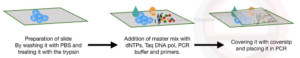
Preparation of Slide
Prepare 2% AES solution just before use
Put 2% AES solution into a Coplin jar & dip glass slides in solution for 60s
Dip slides five times into a different vessel filled with 1,000 ml of distilled water
Air-dry slides in a laminar-flow for a few hours to overnight
Store slides in sealed container at room temperature.
- 250 ml of AES solution is sufficient to treat 200 glass slides. Use slides within 15 d of silanation.
Preparation of Samples
- Starting materials may be cell suspensions; cell cultures on slides; paraffin-fixed tissue; frozen tissue and archived tissue samples.
- Add 10% formalin to the sample for 10-12 hr / overnight.
- Wash the sample/ tissue with PBS twice/ thrice for removal of impurities.
- For cell suspension: Wash with PBS followed by 2 rounds of centrifugation of 25000 rpm, 10 mins.
- For RT PCR, washing with DEPC water.
- Place the tissue or thin film of paraffin embedded tissue to the slide.
- As paraffin can block amplification, treat the tissue with xylene-ethanol (Fresh xylene for 8 mins, then absolute ethanol for 10 mins & air dry)
Proteolytic Digestion
- After fixation, proteolytic digestion (2mg/ml of one proteolytic enzyme used i.e. Pepsin, Trypsin & Proteinase K) followed by washing with alcohol
- Protease digestion is the key step for success of In-Situ PCR, indicated by hole formation in cytoplasmic & nuclear membrane.
- It helps in removal of cross-linked DNA binding protein from nuclear DNA as through creation of holes in the membrane.
- Presence of 10-20 holes in the membrane indicated of proper digestion which is observed under phase-contrast microscope.
In-Situ Amplification
- Apply Master mix on surface of cells/ tissue [Prepare Master mix using one of the 3 primers i.e.
- Fluorescently labelled primer (for Real-time PCR),
- Random hexa / octa nucleotide primers,
- Oligo (dT) primers].
- Cover the slide with coverslip & placed in thermocycler for amplification.
Detection:
- After amplification, PCR products visualized by ISH with a labeled probe (indirect) or direct immunohistochemical detection of labeled nucleotide.
- Direct method may give false result so indirect methods are mainly used & is very sensitive.
- False result due to nonspecific incorporation of labeled nucleotide in to fragmented DNA & in case of aldehyde fixation.
Applications
- Embryogenesis
- Organogenesis
- Detection & diagnosis of viruses & other infectious agents in specific cell types w/i tissues.
- Detection & diagnosis of genetic mutations in inherited diseases.
- Detection of gene & gene expression in a tissue.
- Detection & characterization of tumour cells w/i tissue.
Advantages
- Helps in detection of low copy number of DNA
- With high sensitivity
- Faster assay with shorter turn-around time
- No need of radioactive chemicals
Limitations
- The chance of non-specific binding is higher during in-situ PCR.
- The precision of reaction is low.
- Multiplex is not possible due to the fewer copy number
Conclusions
The in-situ PCR or the slide PCR technique is best method for detection of low copy number of nucleic acid. In reverse transcription in-situ PCR the mRNA is the target from which cDNA is generated and the gene expression level is directly measured on the surface of a slide.
References
- Gerard J.N. 1995. In Situ PCR: Protocols and Applications., Department of Pathology, State University of New York at Stony Brook, Stony Brook, New York.
- Lidonnici, K., B. Lane, and G.J. Nuovo. 1995. A comparison of serologic analysis and in situ localization of PCR-amplified cDNA for the diagnosis of hepatitis C infection. Diagn. Mol. Pathol. (in press).
- Nuovo, G.J. 1994. PCR in situ hybridization: Protocols and applications, 2nd ed. Raven Press, New York.
- Omar Bagasra. 2007. Protocols for the in-situ PCR-amplification and detection of mRNA and DNA sequences 2, NATURE PROTOCOLS.