Wildlife Necropsy: A Practical Guide to Postmortem Examination and Disease Investigation
Kaustubham Narayan Dixit*, Dharam Prakash Shrivastava1
*Undergraduate Student (B.V.Sc. & A.H.), 1Assistant Professor
College of Veterinary Science and Animal Husbandry
Acharya Narendra Deva University of Agriculture and Technology Kumarganj, Ayodhya
Corresponding Author: kaustubham10@gmail.com*, dr.dpsrivastava56@gmail.com1
Abstract
Wildlife necropsy is an important diagnostic tool for determining the cause of death, investigating diseases, and supporting wildlife conservation. This article provides a practical overview of wildlife postmortem examination, highlighting its objectives, major steps, safety precautions, and challenges faced by field and zoo veterinarians in India.
Keyword
Wildlife Necropsy,Postmortem Examination,Disease Diagnosis,Wildlife Conservation,Veterinary Forensics
Introduction
Postmortem examination provides firsthand information on what really happened along the course of the disease. It is an important diagnostic tool and supports other procedures performed in the diagnosis of a disease in a herd or flock (Shrivastava et al., 2025). It is an indispensable, intelligent and scientific tool to diagnose disease, by which the morphological changes in tissues and organs are related to specific causes. Disease diagnosis in wild animals is a difficult task, except during an epidemic. Wild animal disease may occur as an outbreak with high morbidity or mortality or as chronic and protracted conditions. (John et al., 2002)
The objective of this article is to highlight the importance of postmortem examination, challenges faced by field veterinarians as well as zoo veterinarian in India and to guide veterinarian on precautions to be taken along with link to available resources on postmortem techniques, steps in postmortem examination, determination of time of death, postmortem lesions of important diseases etc.
Objective
- To ascertain the exact cause of death.
- To provide valuable insight for clinicians.
- To prevent or mitigate diseases in livestock.
- To provide evidence for potential prosecution in animal-abuse cases.
- To recognize and identify emerging disease or outbreaks of known disease.
- To advance scientific knowledge in various areas of research and development.
- To provide early recognition of disease with zoonotic potential.
Tracing the Past: The Animal’s History
History can help direct the exam and help the person conducting it interpret their findings and any test results. If you have carefully recorded resident observations and maintained your resident’s health records, this will make the process easier. You may be able to provide these records to the relevant veterinarian, or you can use this information to fill in the history portion of the submission form to help give context to the post-mortem exam.
Steps of history taking
- Place or geographical location from where it was recovered.
- Latest weather conditions – drought, flood, storm etc. and ambient temperature.
- Common name, zoological name, sex, approximate age and number of animals died.
- History of the case, duration of illness, morbidity and mortality rate.
- Weight of the body of dead animal (approximate) – if possible length, girth and other body measurement as required.
- Mode of transportation of carcass.
- Date and time of death or found dead.
- Nutritional status of the dead animal.
- Signs of struggle or other evidence relating to death.
- Wounds or other signs of predation.
- Presence of external parasites – ticks, mites, fleas etc.
- Fracture, broken or missing teeth, horns, tusk, bruise, bleeding etc. (John et al., 2002)
Points should be noted during history taking
- Confirmation of death prior to postmortem is must.
- Written consent of owner for doing the postmortem is a must. Requisition letter from competent authority (e.g. Police officer not below rank of inspector or magistrate) is particularly needed for Vetro legal and insurance related cases.
- Recording details of animal identification (e.g. Ear tag, breed, sex, age, length, height, girth behind fore limb, distance between horn tips) and maximum
- possible history (including date and time of death) prior to postmortem is of prime importance.
- Postmortem should be done only when carcass is presented as whole body (In case of drowned animals’ carcass is to be taken out of water and presented for postmortem)
- The carcass should be presented in its actual site as mentioned in the letter from owner/Police.
- In case of any scheduled or endangered animal species, due consent, information/permission should be taken from local forest officials and/or police before proceeding for postmortem examination. (Rath. et al., 2016)
Safety and Biosecurity
The veterinarian, who conducts the post-mortem should protect himself by wearing mask and other protective clothing to safeguard himself from zoonotic diseases like Rabies, Anthrax, Tuberculosis, Psittacosis etc. If the blood smear from ear vein or smear from throat swelling reveals anthrax bacillus, post-mortem should not be conducted on the carcass, since the organisms are aerobic spore formers (The spores can survive as long as 18 years) (John et al., 2002). Scientific disposal of carcass after postmortem: As per Government of India guidelines, it is the responsibility of the veterinarian to ensure the proper disposal of carcass after postmortem examination either by burial or incineration method. It is worthy to remember that the pit/burial site should be 250 meter away from any drinking water source and size of the pit should be bigger than the size of the animal, allowing all parts of the carcass to be buried completely in the pit. A layer of 5 cm lime shall be put at the bottom of the burial pit and again over the carcass before filling the soil. Disposal by incineration should be continued till the animal carcass is reduced to ashes. PM should not be conducted in carcass suspected to have died from anthrax (After suspecting the presence of anthrax bacilli in blood smear from ear vein or oozing out of unclotted blood)
- Postmortem should not be done on carcass that are completely putrefied with liquefaction of all organs.
- Postmortem area should be clean and the chances of environmental contamination should be minimal.
- All natural orifices should be closed with towel or cotton wool soaked in a disinfectant solution before removing carcass to place of postmortem examination.
- Handlers should protect themselves with gloves, apron, mask, cap, gumboot etc.to reduce the contaminations and infections. Movement of people to postmortem site should be restricted (Rath et al., 2016).
Unravelling the Cause of Death: A Step-by-Step Wildlife Necropsy
General guidelines
- Form a team to perform necropsy in advance.
- Mobilize the team members quickly as and when required.
- Include Veterinary Anatomists, Veterinary Pathologists and Wildlife Veterinarians, from nearby Universities.
- Arrange for standard large animal necropsy instruments.
- Assign specific tasks to team members and conduct post mortem in an organized and systematic way.
- Allocate each team different regions like head, foreleg, hindleg abdomen etc.
- Record all data properly.
- Record all body measurements as required.
- Confirm Identity
- PM should be conducted in sufficient Day Light
External Examination
- The first part is the gross (i.e., comprehensive) examination. During this part of the exam, your veterinarian or a veterinary pathologist will perform an external examination.
- Describe any external findings and note the degree of autolysis. If ocular disease is suspected,
- fix the eyes whole using a rapid fixative (e.g. Bouin’s fixative).
- Examine the ears, legs remove them intact
- Examine the nails and sole for lesions.
- Look for the evidence of trauma
Note: The image is illustrative only; it shows a domestic animal, as the basic postmortem procedure is similar in wild animals.
Opening of Carcas
- Make the appropriate midline/ventral incision according to the species.
- Reflect the skin and expose the underlying muscles.
- Examine the subcutaneous tissue and superficial lymph nodes.
- Open the thoracic cavity carefully.
- Examine the pleural cavity for fluid, adhesions or abnormal displacement of organs (John. et al., 2002).
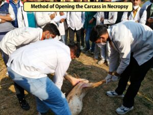
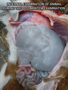
Note: The image is illustrative only; it shows a domestic animal, as the basic Postmortem procedure is similar in wild animals.
Systemic Internal examination
Systematic internal examination means examining the internal organs in a fixed, orderly sequence, so that no organ or lesion is missed. The CZA guide recommends examining body cavities and viscera systematically and collecting appropriate samples for laboratory investigation.
External examination → Thoracic cavity → Respiratory system → Heart & blood vessels → Abdominal cavity → Liver & spleen → Gastrointestinal tract → Urinary system → Reproductive system → Lymph nodes → Brain & spinal cord → Musculoskeletal system → Sample collection & preservation. (John et al., 2002)
The second part is the histopathological examination. During this portion of the exam, tissue samples that were collected during the first part of the exam are examined microscopically. If your primary veterinarian has conducted the first part of the exam, they may submit samples to a diagnostic lab for histopathology. The final part of the process is ancillary, or supportive, testing, as directed by the pathologist or requested by you or your veterinarian. This may entail nutritional testing, testing to detect potential toxins, or testing to detect infectious agents (for example, bacterial cultures, virus isolation, titter testing, etc.)
Note: The image is illustrative only; it shows a domestic animal, as the basic postmortem procedure is similar in wild animals.
Challenges Faced by Field and Zoo Veterinarians During Wildlife Postmortem Examination:
Wildlife necropsy is more challenging than routine domestic-animal postmortem examination because the veterinarian often has limited history, difficult working conditions, unusual anatomy, decomposition, and biosafety risks.
- Limited or Unknown History
- Difficult Field Conditions
- Decomposed Carcasses
- Wild animals are often discovered several hours or days after death.
- Decomposition can destroy or alter important pathological lesions.
- Autolysis may make histopathological and microbiological diagnosis difficult.
- A decomposed carcass should not be considered diagnostically useless. Although decomposition limits interpretation of gross lesions, histopathology and microbiology, valuable evidence may still be obtained from persistent traumatic lesions, foreign bodies, parasites, skeletal injuries, toxicological material and selected tissues. The degree of decomposition and its effect on diagnostic confidence should always be documented.
- Risk of Zoonotic Diseases
- Risk of Physical Injury
- Lack of Diagnostic Facilities
- Difficulty in Sample Collection and Preservation
- Species-Specific Anatomical Differences
Different type of necropsy for PM-examination By Wild and Zoo Vet’s
Where no necropsy is conducted i.e. Anthrax
Partial Necropsy i.e. Rabies
Complete through necropsy
Cosmetic Necropsy i.e. Examination of carcass is done with very less manipulation
Wildlife forensic – A new area of veterinary science
Wildlife forensic – a new area of veterinary science, aids the wildlife. conservation officer in solving crimes against wildlife resources. The illegal killing or taking of wildlife species specimens for commercial purposes has been a major problem throughout the world. Enforcement of conservation regulation is an important tool in the management of wildlife resources an important function of the forensic pathologist is to document the manner, mechanism and cause of death. The causes of death may be an injury or disease that produce a physiological derangement in the body, which result in death. e.g. a gunshot wound to the chest. It may consider such factors as how far the animal travelled or how long it lived after being shot, since they may have direct application in providing the guilt or innocence of the accused. Hence as proof, the Vet should take a photograph of the gunshot wound if any, grossly Of the carcass with the surrounding environment as well as a close-up view of gunshot wound or the injury sites as proof later. In case of gunshot wound, the skin with the wound site should be taken and preserved for future as evidence in the court (John et al., 2002)
Conclusion
Wildlife postmortem examination is an essential tool for determining the cause of death, identifying diseases, supporting conservation efforts, and providing valuable forensic evidence. A systematic approach—from obtaining the animal’s history and conducting a careful external examination to internal examination, appropriate sample collection, preservation, and laboratory investigation—is essential for obtaining reliable results. Wildlife veterinarians must also consider species-specific anatomy, decomposition, field conditions, zoonotic risks, and legal requirements. Proper documentation, biosafety, and timely examination can greatly improve diagnostic accuracy and contribute to wildlife health, disease surveillance, and conservation.
REFERENCES
- Shrivastava, D.P., Niyogi, D. and Tripathi, K.K. (2025). Laboratory Manual on Veterinary Pathology.
- John M. C., Manohar B. M., Ramadass P., Jaya Thangaraj M.G. (2002). Central Zoo Authority (CZA), Government of India. Basic Postmortem Requirements for Zoo Veterinarians. Central Zoo Authority, Ministry of Environment & Forests, Government of India.
- Hess, Tara. Understanding the Potential Benefits of Post-Mortem Examinations for Sanctuary Residents.
- Rath P.K., Mishra B.P., Mishra J. (2016). Vet Helpline India. Postmortem Diagnosis and Field Veterinarians in India. Vet Helpline India. Available at: vethelplineindia.co.in/postmortem-diagnosis-and-field-veterinarians-in-india.
|
Submitted By: Kaustubham Narayan Dixit BVSc & A.H-IIIrd Year Collage of Veterinary Science and Animal Husbandry Acharya Narendra Deva University of Agriculture and Technology |
Dr.D. P Shrivastava Assistant Professor Department of Veterinary Pathology Collage of Veterinary Science and Animal Husbandry Acharya Narendra Deva University of Agriculture and Technology
|