Blood in Milk in Bovines: Causes and Available Treatment Options
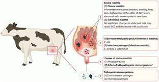
Blood in milk or hemogalactia or hemolactia produces milk that is reddish or pinkish due to the presence of blood. Hemogalactia causes economic losses because bloody milk is rejected by the industry and consumers. It is common in cows after parturition. Blood in milk occurs from 2-8 days after parturition. Blood in milk is usually diagnosed based on the clinical signs. Trauma to teat and udder is one of the common causes of blood in milk. Bacteria (Leptospira spp., Brevibacterium erythrogenes, Serratia marcescens, Micrococcus cerasinus, Micrococcus chromidrogenes rubber, Micrococcus roseus, Lactorube faciens gruber, Sarcina rubra etc.), some viruses and red yest (Monascus purpureus) may cause systemic infection associated with intravascular hemolysis and capillary damage in udder leading to reddish or pinkish discoloration of milk . Leptospirosis is one of the common causes of blood in milk in dairy cows. In leptospirosis, milk from all the quarters would be red in color, thick in consistency and it contains both blood and milk clots . Cattle affected with hemogalactia are characterized by low platelets count and it may show pinkish or reddish discoloration of milk due to leakage of blood into milk . Blood in milk or hemogalactia in lactating dairy cattle is common in heifers and multiparous cows . Reddish or pinkish discoloration of milk in cattle observed due to thrombocytopenia that may cause leakage of blood in milk.
The animal husbandry and dairying sector in India contributes about 33% to GDP and thus a promising industry for the growth of the country. Among many constraints to the dairy industry in terms of infectious and metabolic diseases, a condition like Hemolactia (blood in milk) also leads to heavy economic losses to the dairy farmers. Dairy farmers and farmers with one or two animals frequently seek the veterinary help for the treatment of cows and buffaloes with hemolactia condition. Cases of the condition are sporadic but several lactating animals can be affected at a time. This condition adds to economic losses as milk have to be discarded due to rejection by consumers due to its abnormal color and also the treatment cost. So, the knowledge of etiology and cheap and effective treatment strategies can help the farmers to cut down the economic losses.
CAUSES
There can be multiple causes for this condition. A few important causes are as below:
1. Physiological Hyperemia and Hemorrhage: Hyperemia (increased blood supply to an organ/tissue) of the mammary gland which is occasionally seen at late gestation and for a short period just after parturition. Blood in milk due to this state normally persists no longer than 14 days at the most. However, if such udder is not milked completely can precipitate the condition at any stage of lactation. Hemorrhage by diapedesis (passage of blood cells through capillary walls into the tissues), is quite common just after calving or as sequalae to physiological hyperemia. Hemorrhage due to diapedesis can occur at any stage during the lactation. The color of the milk can be slight pinkish tinged to red depending upon the severity of hemorrhage. Harsh milking by hand or machine may result in hemorrhage due to epithelial damage. Trauma to udder and teat is also one of the common causes of blood in milk due to hemorrhage. If hemorrhage is due to rupture of major vein of udder then frank venous blood like secretion can be there in milk.
- Systemic infections: Several bacterial infections including Leptospira species, Brevibacterium erythrogenes, Serratia marcescens, Micrococcus species, Lactorubefaciens gruber, Sarcina rubra etc., along with some viruses and red yeast like Monascus purpureus may cause systemic infections which can lead to intravascular hemolysis and capillary damage in udder and causes pinkish to reddish discoloration of milk. Among systemic infections, Leptospirosis is one of the common causes of blood in milk in dairy animals. Red colored milk with thick consistency from all four teats may or may not be accompanied with blood and milk clots is characteristic of Leptospira infection. Soft udder and cold mastitis (mastitis with no sign of inflammation) is another characteristic clinical feature of leptospiral mastitis which develop after some nonspecific signs of fever, decreased milk yield, hemoglobinuria etc.
3. Natural toxins or dyes: Sometimes toxins from plants viz; conifers, poplars, alders, ranunculi etc. (shown in fig.1) may cause capillary damage leading to reddish discoloration of milk. Moldy sweet clover (dicoumarin poisoning- anticoagulant) can cause bloody milk. Sometimes dye containing leafy plants can cause reddish discoloration of milk without any pathological condition
4. Deficiency of blood platelets (Thrombocytopenia): Cattle with diseases where low platelet count occurs as one of the manifestation, may show reddish or pinkish discoloration of milk due to leakage of blood into milk.
5. Acute or chronic mastitis: In chronic mastitis, due to harsh milking, straining of tissue or due to lying on uneven surfaces can cause temporary or protracted hemorrhage into the milk due to rupture of vascular granulation tissue.
6. Miscellaneous causes: Vitamin C deficiency, penetrating lacerations, whip injuries, horn injuries.
Treatment Options Available for Hemolactia in Bovines
Treatment strategies—
- Calcium borogluconate Intravenous 300-450 ml (for 2-3 day). Calcium has a coagulant effect.
- Coagulants Parenteral/ Local Tranexamic acid, 500 mg/ml; 10-15ml i/m bid · Etamsylate, 250 mg/ml; 5-10 mg/kg b. wt. i/m . · Adrenochrome monosemicarbazone, 5 mg/ml, i/m (50mg total)
(Local application/intramammary is considered as more efficient than parenteral in severe cases.)
- Vasoconstrictors: Parenteral/Local 5-8 ml (1:1000) epinephrine s/c · 5ml epinephrine+ 20ml Normal saline intramammary · Ergonovine maleate (total 10-20mg) i/m · Methylergometrine hydrogen maleate (total dose 2mg) i/m (Circulatory system of the udder is very sensitive to the vasoconstrictor action of adrenaline.)
- Vitamin C Ascorbic acid injection @ 7.5 mg/kg body weight intramuscular · Ascorbic acid tablets (20-30 tablets each containing 500 mg vitamin C) { Vitamin C has anti-oxidant effect.}
- Antibiotics For leptospiral mastitis: Streptomycin (25mg /kg b.wt. i/m for 3-5 days) or after antimicrobial susceptibility testing for other causes of mastitis. { Intramammary or parenteral antibiotics can be given}
- Vitamin K @ 10 ml i/m for 3 days { Vitamin K is antihemorrhagic}
- Formalin 35% (5ml) + Dextrose saline (500ml) i/v · 10% Formalin (10-30 ml) orally · 0.37% solution of Formalin in normal saline i/v for 3-4 days
- Styplon® Vet bolus 1-2 bolus bid 3-4 days { Silk cotton tree(Shalmali) And Malabur nut (Vasaka) are key ingredients.}
- Camphor 20 parts camphor powder (finely ground) in 80 parts of olive oil) 30-60 ml of camphorated oil i/m { Volatile acids released by camphor act as styptic (Ethno-veterinary Treatment)}
- Homeopathic treatment Homeopathic complex of Phytolacca 200c, Calcarea fluorica 200c, Silicea 30c, Belladona 30c, Bryonia 30c, Arnica 30c, Conium 30c and Ipecacuanaha 30c. 10 pills four times daily until recovery.
- Tonophosphan Vet® @ 10 ml i/m for 3-4 days. { Phosphorus maintains the RBC membrane integrity}
- 200 gram of curry leaves (Murraya Koengii) + juice of 10 normal sized lemons (Citrus limon) p.o. twice daily for 3-4 days.
- 250 grams of turmeric powder in one liter of warm milk + 250 grams of ‘sambaloo’ leaves and giving as a drench for 2-3 days.
SUPPORTIVE TREATMENT AND OTHER MEASURES
1. Application of ice cold water or crushed ice (packed in a cloth) helps in control of hemorrhage through vasoconstriction.
2. The sand hosed with cold water at least four times can be used as bedding material for affected animals, so that when the animal rest on sand, it will lead to vasoconstriction and control of hemorrhage in the udder or teat.
3. The feed suspected for causing blood in milk should be changed.
4. Cleaning of the animal shed and to provide kutcha floor with even surface for animals.
Compiled & Shared by- This paper is a compilation of groupwork provided by the Team, LITD (Livestock Institute of Training & Development)
Image-Courtesy-Google
Reference-On Request


