BY: DR.Manoranjan Nanda, Bhubaneswar.
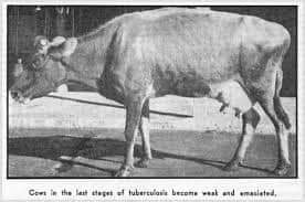
Bovine tuberculosis is a chronic and slowly progressive disease of cattle that emerges periodically in the India. Its incubation time ranges from months to years. Most often, infected cattle will show little to no outward signs of infection. When clinical signs are present, they will often be vague, such as weight loss, depression, and sluggishness. Transmission of tuberculosis between animals occurs when susceptible animals are in close contact with respiratory secretions or aerosols from infected animals. Close contact is necessary for transmission; the infection is not considered to spread easily between cattle separated by any distance.
Tuberculosis is a chronic disease of many animal species and poultry caused by bacteria of the genus Mycobacterium.
It is characterized by development of tubercles in the organs of most species.
Bovine tuberculosis is caused by Mycobacterium bovis. It is a significant zoonotic disease.
🐂Transmission :
An infected animal is the main source of transmission. The organisms areexcreted in the exhaled air and in all secretions and excretions.
Inhalation is the chief mode of entry and for calves infected milk is an important source of infection. When infection has occurred tuberculosis may spread: a) by primary complex (lesion at point of entry and the local
lymph node) and b) by dissemination from primary complex.
#Antemortem_findings:
1. Low grade fever
2. Chronic intermittent hacking cough and associated pneumonia
3. Difficult breathing
4. Weakness and loss of appetite
5. Emaciation
6. Swelling superficial body lymph nodes
TB usually has a prolonged course, and symptoms take months or years to appear.
The usual clinical
signs include:
– weakness,
– loss of appetite,
– weight-loss,
– fl uctuating fever,
– intermittent hacking cough,
– diarrhea,
– large prominent lymph nodes.
However, the bacteria can also lie dormant in the host without causing disease.
#Postmortem findings:
1. Tuberculous granuloma in the lymph nodes of the head, lungs , intestine and
carcass.
These have usually a well defined capsule enclosing a caseous mass with a calcified centre.
They are usually yellow in colour in cattle, white in buffaloes and greyish
white in other animals.
2. Active lesions may have a reddened periphery and caseous mass in the centre of a
lymph node.
3. Inactive lesions may be calcified and encapsulated
4. Nodules on the pleura and peritoneum
5. Lesions in the lungs , liver, spleen, kidney
6. Bronchopneumonia
7. Firmer and enlarged udder, particularly rear quarters
8. Lesions in the meninges, bone marrow and joints
The diagnosis may be confirmed by making a smear of the lesion and with Ziehl-Neelsen. The TB bacterium is a very small red staining bacillus.
#Discussion :
Mycobacteria invade cattle by respiratory (90 – 95 %) and oral routes (5–10 %).
Congenital infection in the bovine fetus occurs from an infected dam.
Tuberculosis lesions can be classified as acute miliary, nodular lesions and chronic organ tuberculosis.
Young calves are infected by ingestion of contaminated milk. The incidence of human tuberculosis caused by Mycobacterium bovis has markedly dropped with the pasteurization of milk.
It also has dropped in areas where programs of tuberculosis eradication are in place.
Man is susceptible to the bovine type. In cattle, lesions of tuberculosis caused by the avian type are commonly found in the mesenteric lymph nodes. Tuberculosis in small ruminants is rare. In pigs the disease may be caused by the bovine and avian types. Superinfection is specific in cattle.
#Judgement :
Carcass of an animal affected with tuberculosis requires additional postmortem
examination of the lymph nodes, joints, bones and meninges.
It is suggested that the Codex Alimentarius judgement recommendations for cattle and buffalo carcasses be followed.
🐂Carcasses are condemned
i. where an eradication scheme has terminated or in cases of residual infection or reinfection
ii. in final stages of eradication – natural prevalence low
iii. during early stages in high prevalence areas
Carcass of a reactor animal without lesions may be approved for limited distribution. If the
economic situation permits, this carcass should be condemned.
Heat treatment of meat is suggested during early and final stages of an eradication programme: in low and high prevalence areas where one or more organs are affected, and where miliary lesions, signs of generalization or recent haematogenous spread are not observed.
If the economical situation permits, then the carcass is condemned.
In some countries, the carcass is approved if inactive lesions (calcified and/or encapsulated) are observed in organs and without generalization in lymph nodes of carcass.
Differential diagnosis :
👉 Lung and lymph node abscess,
👉pleurisy.
👉 pericarditis,
👉chronic contagious pleuropneumonia,
👉actinobacillosis,
👉mycotic and parasitic lesions,
👉 tumours,
👉 caseous
lymphadenitis Johne’s disease,
👉adrenal gland tumour and lymphomatosis.
🐂What is being done to prevent
or control this disease?
The standard control measure applied to TB is test and slaughter. Disease eradication programs consisting of post mortem meat inspection, intensive surveillance including on-farm visits, systematic individual
testing of cattle and removal of infected and in- contact animals as well as movement controls have been very successful in reducing or eliminating the disease.
Post mortem meat inspection of animals looks for the tubercles in the lungs and lymph nodes
(OIE Terrestrial Animal Health Code). Detecting these infected animals prevents unsafe meat
from entering the food chain and allows veterinary services to trace-back to the herd of origin of the infected animal which can then be tested and eliminated if needed.
Pasteurisation of milk of infected animals to a
temperature suffi cient to kill the bacteria has
prevented the spread of disease in humans.
Treatment of infected animals is rarely attempted because of the high cost, lengthy time and the larger goal of eliminating the disease.
Vaccination is practiced in human medicine, but it is not widely used as a preventive measure in animals: the effi cacy of existing animal vaccines is variable and it interferes with testing to eliminate the disease. A number of new candidate vaccines are currently being tested.
Reference:On request.


