CLASSIFICATION OF PLACENTA AND ITS FUNCTION IN DOMESTIC ANIMALS
Renu Yadav, Amit vishen
Department of Veterinary Anatomy, College of Veterinary and Animal Science
G.B. Pant University of Agriculture & Technology
Pantnagar, Uttarakhand, Pincode-263145
Introduction
The term placenta was derived from Greek word it means flat cake. Placenta is a temporary organ. It is a unique organ that develops in mammalians for the development of the fetus. It is an apposition of fetal membranes to the endometrium to permit physiological exchange between the fetus and the mother. It mediates the metabolic exchanges of the developing individual through an intimate association of embryonic tissues and of certain uterine tissues, serving the functions of nutrition, respiration, and excretion. It works as an endocrine gland. It will secretes lactogen, progesterone, etc. hormones. Placental hormones act to adapt maternal physiology to pregnancy and lactation. Highest concentration of trace elements was observed in the region of maternal-fetal interface, a region with trophoblast. Inadequate transfer of essential minerals from mother to fetus results in deficiency in these nutrients in the embryo, causing damage to its growth and abnormalities in metabolism, central nervous system and bone.
Classification of placenta:
- Based on loss of maternal tissue in placental shedding:
Deciduate or conjoined or placenta vera or True placenta – Villi fused with uterine mucosa where uterine lining tears and bleeding seen. The trophoblast invades the endometrium and destroys the superficial endometrial tissue in deciduate placenta. Ex. Cat, rabbit, mouse, women, primates, Insectivores
Non-deciduate type or Semi placenta – Villi of chorionic are in apposition with uterine lining but don’t fuse with it. Trophoblast does not invade with endometrium. Hence there is no shedding of maternal tissue/ breeding. Ex. Cow mare, ewe, dog and pig. In Non-deciduate type, the fetal membranes and placenta are expelled at the time of parturition, leaving the endometrium intact except in ruminants in which only the surfaces of the carcuncles are devoid of epithelium after the caruncles sloughs about 6–10 days following parturition.
(B) Based on foetal membrane involved
Chorionic: when allantois is lacking, it is a rare type
Choriovitelline : in this yolk sac is large and unites with the chorion. This is also called as Inverted yolk sac placentation. ex. Some marsupials.
Chorioallantoic: in all higher mammals including domestic animals allantois comes in to contact and fuses with chorion with vascular villi. Ex. Cow, sheep, goat, horse, women and dog
(C) Based on nature of foetal maternal contact surface
Folded placenta: The both sides (foetal and maternal) are folded ex. Sow.
Villous placenta: The branch of villi fit in to maternal crypts (horse, cow) or freely exposed
to maternal blood (women).
Labyrinthine: The villi are fused to form a labyrinth. Ex Dog, cat, lower rodents, vampire
bat, rabbit .
(D) Based on shape of placenta
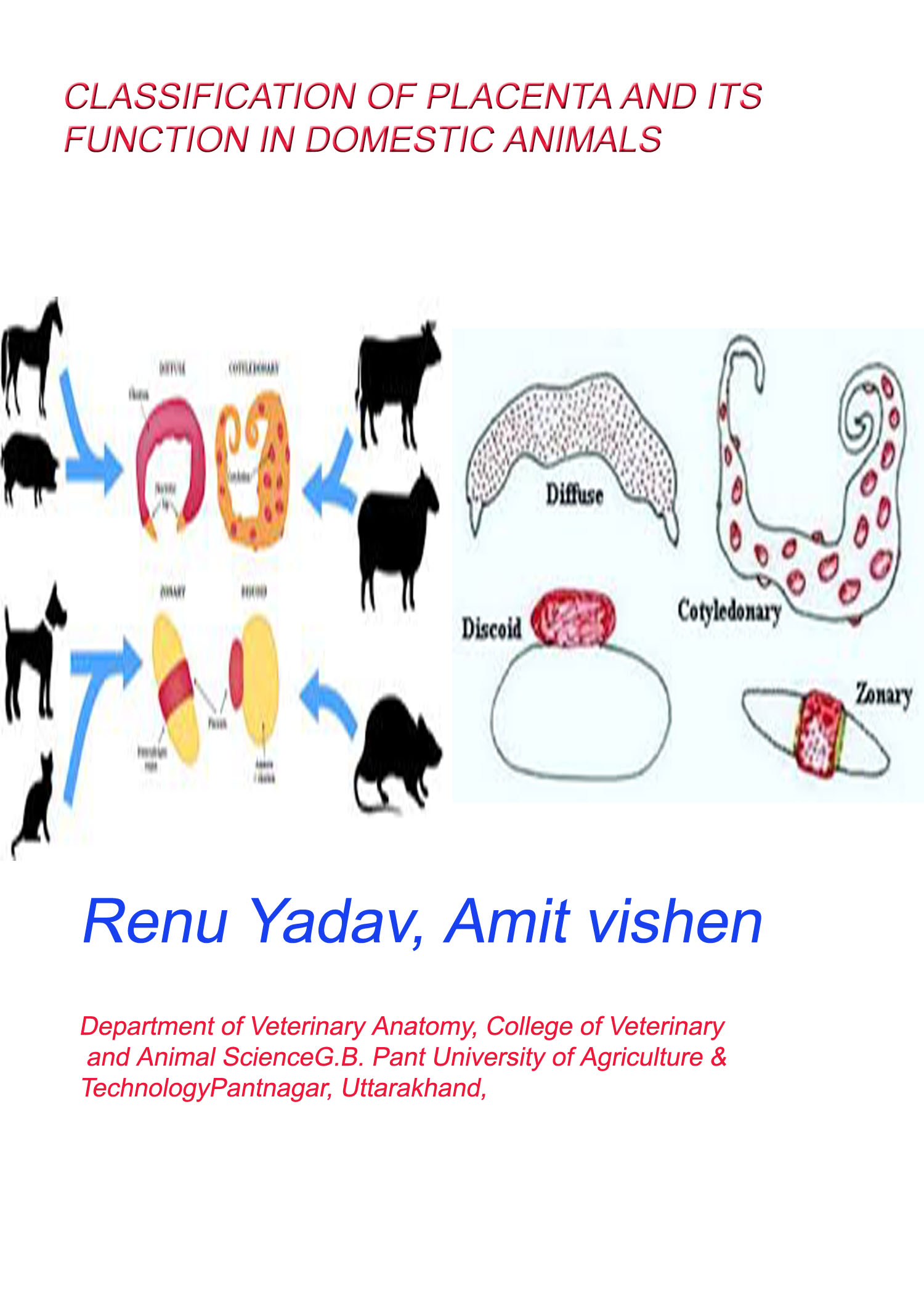
Diffuse placenta: It is found in wide range of species, including pigs, horses, camels,
lemurs, whales, dolphins, kangaroos and possums. The villi of the chorion are distributed
more or less evenly over the entire surface of the chorionic sac.
Cotyledonary placenta: The cotyledonary placenta is characteristic of the ruminants;
instead of being uniformly distributed over the entire surface of the chorion, the chorionic
villi are clumped together in to well developed circular regions known as cotyledons.
These cotyledons develop only in those regions of the chorion that overlie predetermined aglandular areas of the endometrium known as the caruncles. The fetal cotyledon and maternal caruncle unite to form a placentome, and these placentomes are the only sites of maternal-fetal exchange, the intercotyledonary chorion being devoid of villi and unattached to the endometrium. There are three basic shapes to placentomes. The convex type is typical of cattle and often has a narrow base giving it a mushroom shape. Flat placentomes are found mostly in deer, and the concave type is found in sheep and goats. The sheep placentome has a central concavity where extravasated maternal erythrocytes are taken up and processed by columnar trophoblast cells
Zonary Placenta: It is characteristic of the carnivores, and is the result of an aggregation of chorionic villi to form a band that encircles the equatorial region of the chorionic sac. It may be complete, as in dog and cat, or incomplete, as in bears, seals and mustelids.
(E) BASED ON STRUCTURAL LAYERS BETWEEN MATERNAL AND FETAL BLOOD
Epithelio chorial placenta : Ex Pig, Horse, (Ungulates Lemmures)The foetal chorion is in contact with epithelium of the uterus hence it is called epithelio-chorial placenta. In between foetal, maternal parts six layers are present. If all the six layers are present the placenta is called epithelio-chorial placenta.
Syndesmochorial (Ruminants): All tissues of the previous type are present with the exception of the maternal epithelium. Due to loss of uterine epithelium, chorionic epithelium contacts vascular connective tissue of mother.
Endotheliochorial (Dog and Cat): The chorion of the foetus will come in contact with the endothelium of mother ‘s uterus, hence it is called endothelio-chorial placenta.
| Species | Classification of Chorioallantoic Placentas | ||
| Chorio Villous Pattern | Maternal-Fetal Barrier | Loss of Maternal Tissue at Birth | |
| Pig | Diffuse | Epitheliochorial | None (nondeciduate) |
| Mare | Diffuse and Microcotyledonary | Epitheliochorial | None (nondeciduate) |
| Sheep, goat,
cow, water buffalo |
Cotyledonary | syndesmochorial | None (nondeciduate) |
| Dog, cat | Zonary | Endotheliochorial | Moderate (deciduate) |
| Man, monkey | Discoid | Hemochorial | Extensive (deciduate) |
Function of the Placenta
Respiration
The placenta is the only source of oxygen for the fetus. Fetal haemoglobin has a higher affinity for oxygen than adult haemoglobin. The fetal haemoglobin is more attractive to oxygen molecules than the maternal haemoglobin. As a result, when maternal blood and fetal blood are nearby in the placenta, oxygen is drawn off the maternal haemoglobin, across the placental membrane, onto the fetal haemoglobin. Carbon dioxide, hydrogen ions, bicarbonate and lactic acid are also exchanged in the placenta, allowing the fetus to maintain a healthy acid-base balance.
Nutrition All of the nutrition for the fetus comes from the mother. This nutrition is mostly in the form of glucose, which is used for energy and growth. The placenta can also transfer vitamins and minerals to the fetus.
Excretion The placenta performs a similar function to kidneys, filtering waste products from the fetus. These waste products include urea and creatinine.
Endocrine The major hormones of ruminant placentae are progesterone and other progestins, estrogens and placental lactogen.The sheep placenta produces enough progesterone that by roughly day 70 the corpora lutea can be removed and pregnancy will not be interrupted. In contrast, luteal progesterone is required throughout gestation in cattle and goats because their placentae secrete much smaller quantities of progesterone. In reality, a large amount of progesterone is synthesized by the goat placenta, but most is converted to a biologically inactive pregnane before secretion. The patterns of placental lactogen secretion is quite different in cattle and sheep. The bovine hormone is detected in maternal serum at about 4 months of gestation and remains low through parturation. Both dogs and cats produce the hormone relaxin during pregnancy.
In pregnant bitches, relaxin is first detected in serum about 4 weeks into gestation, and increases substantially during the remainder of gestation. The placenta is known to be the primary site of secretion of relaxin in dogs, although a smaller contribution is made by the ovaries and luteal synthesis of relaxin persists for several weeks after parturation. Relaxin is not present in serum of pseudopregnant bitches, and thus can be reliably used as a pregnancy test. Equine chorionic gonadotropin (eCG): This glycoprotein hormone is secreted by a part of the fetal placenta called endometrial cups. In pregnant mares, eCG is usually first detectable in blood between 35 and 40 days of gestation, rise rapidly to peak about 60 days, then decline slowly to become undetectable at about 120 days of gestation. The high blood levels of eCG during this time stimulate development of ovarian follicles. Equine CG is also known as pregnant mare’s serum gonadotropin or PMSG.
Conclusion: Placenta is an apposition of fetal membranes to the endometrium to permit physiological exchange between the fetus and the mother. It mediates the metabolic exchanges of the developing individual through an intimate association of embryonic tissues and of certain uterine tissues, serving the functions of nutrition, respiration, and excretion. It works as an endocrine gland. It will secretes lactogen, progesterone, etc. hormones. Placental hormones act to adapt maternal physiology to pregnancy and lactation.


