Clinical Management of Postpartum Uterine Prolapse Concurrent with Milk Fever
Sudhanshu Pratap Singh1, Ankesh Kumar2, Nitu Sourya3, Mukesh Sahu4 and Avaneesh Kumar Singh 5
1 M. V. Sc. Scholar Department of Veterinary Gynaecology & Obstetrics, BASU, Patna 14
2 Department of Veterinary Clinical Complex, BVC, Bihar Animal Sciences University BASU, Patna, Bihar, India
3 BVC, Bihar Animal Sciences University (BASU), Patna, Bihar, India
4Department of Veterinary Gynaecology and Obstetrics, GBPUAT – Pant Nagar, Uttarakhand, India
5Department of Veterinary Gynaecology and Obstetrics, DUVASU, Mathura, Uttar Pradesh, India
*Corresponding author: – Email: spsingh91292@gmail.com
Key words: Uterine prolapse, milk fever, hypocalcaemia
Running title: Clinical Management of Postpartum Uterine Prolapse Concurrent with Milk Fever
Abstract
Uterine prolapse is a non-hereditary complication associated with calving that occurs immediately after parturition in most of the cases and occasionally up to several hours afterwards. Although, the predisposing factors like excessive oestrogen content in the feed, hypocalcaemia, increased straining, poor uterine tonicity, forced extraction of the foetus, weight of retained foetal membranes, conditions that increased intra-abdominal pressure including tympany are reported for uterine prolapse. A 4 years old crossbreed cow was attended at the farmer’s doorstep for treatment of uterine prolapsed which was noticed by the farmer after 4 hrs of parturition. On clinical examination, it was noticed that the cow was in sternal recumbence and unable to stand, subnormal body temperature, cold skin and extremities, suspended rumination, defecation, urination and ocular mucous membrane was congested. Prolapsed mass was found to be lying on the ground and the prolapsed uterus mass was swollen, oedematous, partially necrotic and stained with dung materials and debris. The Animal was showing signs of discomfort, restlessness, continuous straining. Based on the clinical examination, it was diagnosed to be a case of postpartum uterine prolapse concurrent with milk fever. Epidural anaesthesia was achieved by infiltration of 7 ml lignocaine hydrochloride (2%) into the first sacrococcygeal vertebral space to prevent straining during replacement of the prolapsed organ. The prolapsed uterus was washed thoroughly with a mild antiseptic solution (1:1000 potassium permanganate) for removal of the necrotic tissue, debris and dung material. The foetal membrane was removed carefully from the maternal caruncles. The prolapsed mass was properly repositioned, and suture was applied. Inj. Hydroxy progesterone (Duraprogen) @2ml IM was given to compete against the oestrogen level. For post operative management, Inj. Amoxycillin sodium and Sulbactam sodium (Amoxirum Forte 3g) @ 10mg/kg BW IM was applied for 5 days to overcome infection and Flunixin meglumine (Megludyne inj.- 10 ml IV) for palliative management. Inj. Chlorpheniramine maleate (10ml IM) as antihistaminic, Calcium borogluconate (450ml slow IV) and Rintose (1000ml IV) was applied. The animal has stopped straining and has started feeding and drinking normally after 12 hours the suture was removed on 7th day.
Key words: Uterine prolapse, milk fever, hypocalcaemia
Introduction
Uterine prolapse is a non-hereditary complication associated with calving [1] that occurs immediately after parturition in most of the cases and occasionally up to several hours afterwards. It is observed most commonly in cow and ewe, occasionally in sow and rarely in dogs, cats and mare [2]. Prolapse of uterus mostly seen in pupiparous dairy cows. In milk fever the atonic uterus may prolapse possibly due to the increased abdominal pressure of labour [2]. In crossbred cattle, prolapse of uterus is usually associated with hypocalcaemia or milk fever [3]. Although the predisposing factors like excessive estrogen content in the feed [3], hypocalcemia, increased straining [4], poor uterine tone, forced extraction of the foetus, weight of retained fetal membranes, conditions that increased intra-abdominal pressure including tympany are reported the exact etiology of uterine prolapse is still unclear [5]. Milk fever is a metabolic disease of mature dairy cows that occur just before or soon after calving, characterised by hypocalcemia, severe muscular weakness, sternal and lateral recumbency. The aim of this case report is to highlight the treatment and management of uterine prolapse concurrent with milk fever of a crossbred cow in field condition.
Case History and Clinical Observation
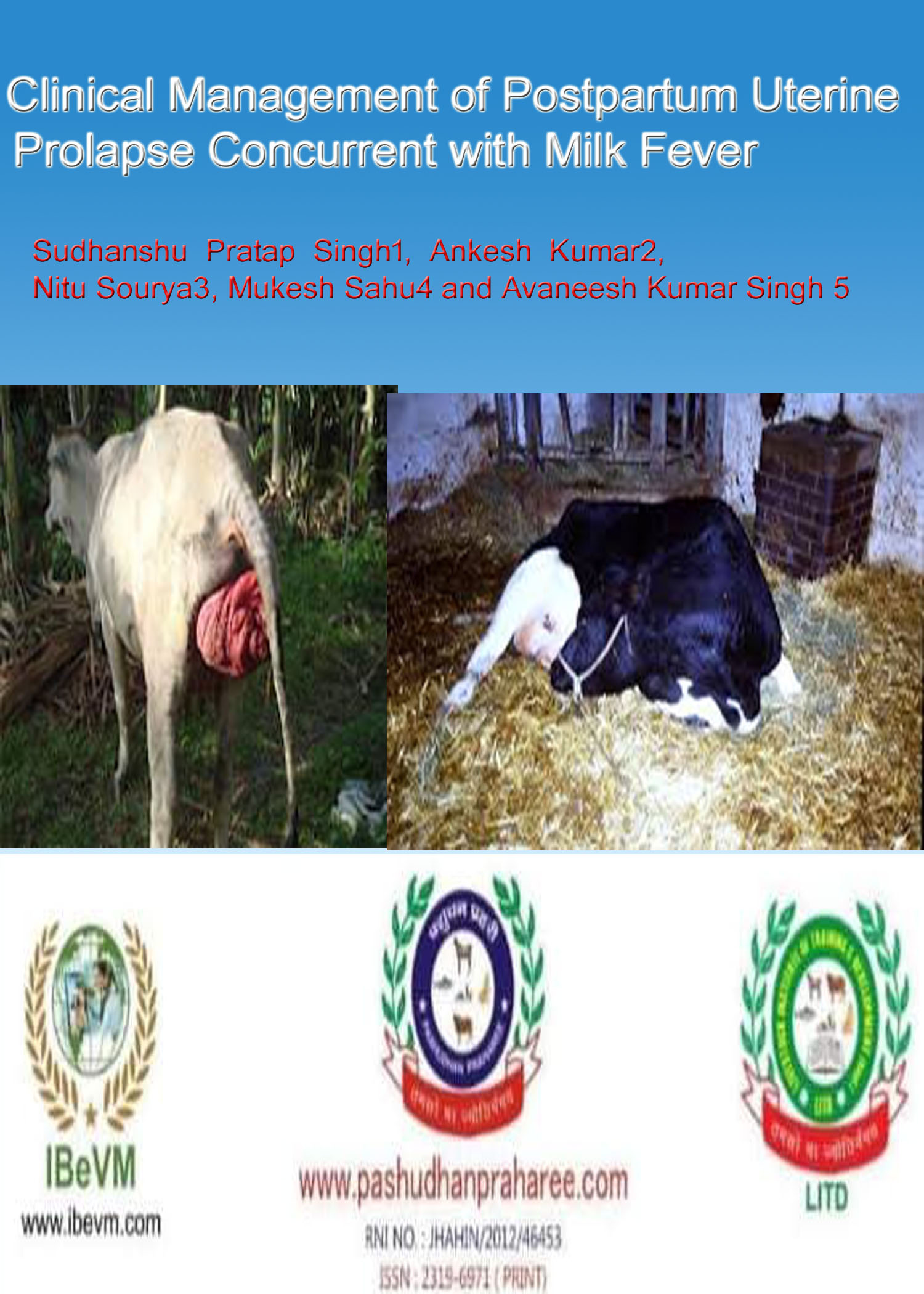
A cross breed cow (4 years) was attended at the farmer’s doorstep for treatment of uterine prolapse concurrent with milk fever which was noticed by the farmer after 4 hrs of parturition. On clinical examination, it was noticed that the cow was in sternal recumbency, animal was unable to stand, subnormal body temperature, cold skin and extremities, suspended rumination, defecation, urination and ocular mucous membrane was congested. Prolapsed mass was found to be lying on the ground and the prolapsed uterus mass was swollen, edematous, partially necrotic and stained with dung materials and debris. The Animal was showing signs of discomfort, restlessness, continuous straining. Based on the clinical examination, it was diagnosed to be a case of Uterine prolapse with milk fever.
Laboratory Diagnosis
Blood was collected from jugular vein for biochemical evaluation. Serum biochemistry results showed hypocalcemia 5.6 mg/ dl.
Clinical management
Epidural anesthesia was achieved by infiltration of 7 ml of 2% lignocaine hychlochloride into the first sacrococcygeal vertebrae to prevent straining during replacement of the prolapsed organ. The prolapsed uterus was washed thoroughly with a mild antiseptic solution (1:1000 potassium permanganate) for removal of the necrotic tissue, debris and dung material. The foetal membrane was removed carefully from the maternal caruncles . At first the ventral portion of the prolapsed part was replaced followed by the dorsal part. The moderate force was applied on prolapsed mass to reduce manually. The prolapsed mass was properly repositioned and suture was applied to the exterior. The case was managed by injecting Hydroxy-progesterone (Duraprogen, Vetcare) @2ml IM to compete against the oestrogen level. For supportive therapy 1000ml of Rintose was infused and an antibiotic course of amoxycillin and salbactum @ 10 mg/kg body weight (Amoxyrum Forte 3 gm, Virbac) IM for 5 days was
administered, Flunixine meglumine 10 ml iv @ 1.1- 2.2 mg/kg (Megludyne, Virbac) to reduce the pain and inflammation.
antihistaminic (inj.Chlorpheniramine maleate)10ml i/m, and Mifex (Calcium borogluconate) @ 450 ml slow i/v After about 12 hours the owner reported that the animal has stopped straining and has started eating and drinking normally. The case was followed up for the next 5 days and the suture was removed on 10th day.
Results and Discussion
Prolapse of the uterus is a common complication in high yielder cows at the time of foetus expulsion. Since hypocalcemia is the most common cause of uterine prolapse [7]. Calcium borogluconate therapy was given in this case with management of prolapsed mass. To prevent secondary bacterial infection an injectable broad-spectrum antibiotics were administered for 5 days after repositioning of prolapsed uterus mass on its normal anatomical position . Early intervention is required. Delayed intervention usually results to poor prognosis of the case due to the risk of hemorrhage, shock and death (Andrews et al., 2008) [8]
Conclusion
Lowering dietary calcium levels during dry period is very important for prevention of milk fever, as well as to balance the acid-base diet ratio; Dietary Cation-Anion Difference (DCAD) [6]. Incomplete milking must be done after calving for 2-3 days. Avoide the estrogenic feed & fodder to prevent the prolapse. Case of postpartum prolapse with milk fever in cow can be treated with manual pressure followed by administration of calcium therapy along with other supplemental therapy for the successful management of the case.
References
- Kumar AS, Yasotha A. Correction and management of total uterine prolapse in a crossbred cow. Journal of Agriculture and Veterinary Sciences. 2015; 8(1):14-16.
- Roberts SJ. Veterinary Obstetrics and Genital diseases, 2nd edn. C.B.S. Publisher and distributors, Delhi, 1971, 308-313.
- Kumar AS, Yasotha A. Correction and management of total uterine prolapse in a crossbred cow. Journal of Agriculture and Veterinary Sciences. 2015; 8(1):14-16.
- Roberts SJ. Veterinary Obstetrics and Genital Diseases (Theriogenology). 2nd ed. Reprint, C.B.S. Publisher and distributors, Delhi, India, 2004, 300-40.
- Noakes ED, Parkinson TJ, England GCW. Post parturient prolapse of the uterus. In: Arthurs Veterinary Reproduction and Obstetrics. 8th ed. Harcourt (India) Pvt. Ltd., New Delhi, 2001, 333-338.
6.DeGaris PJ and Lean IJ (2009). Milk Fever in Dairy Cows: A Review of Pathophysiolgy and Control Principles., The Vet. J. 176: 58-69
- Arthur GH. Veterinary Reproduction and Obstetrics.8th ed. Harcourt (India) Private Limited, 2001.
- Andrews AH, Blowey RW, Boyd H and Eddy RG (Eds.).(2008). Bovine medicine: diseases and husbandry of cattle. John Wiley and Sons. pp. 514-515.
.


