COMMON GENETIC DEFECTS IN DOMESTIC ANIMALS
The terminology concerning defects or abnormalities in cattle can render confusion if there is not a distinction between the adjectives congenital and hereditable. The word congenital is derived from the Latin words con and genitalis. Con means with or together and genitalis means to beget or reproduction. Thus the word congenital describes those conditions which are present at birth as a result of the developmental process. Heredity is dervied from the Latin word hereditas or heirship. Thus hereditable indicates those conditions in the young which are present as a result of parental genotypes. Most, but not all inherited developmental defects are apparent at birth and therefore can be said to be congenital. However, not all congenital defects can be accurately termed hereditable.
An illness caused by inborn abnormalities in genes or chromosomes • Caused by vertical transmission of defective genes to the offspring • Genetic defects are of minor concern in production, but increase in frequency and number of carrier animals of these alleles/defects may cause significant loss in the industry
Some of the defects are as follows-
1.Arthrogryposis Multilpex (Curly Calf Syndrome)
Generally found in Angus cattle • Disease is due to deletion of a section of DNA (at least 38,000bp) • this defect is an autosomal recessive trait • no protein is produced due to the deletion • Main clinical symptoms are: bilateral tibia (twisted rear leg with anchylosed joints) Abdominal hernia and cranial defect (cranioschisis with meningocoele)
2.Syndactylism (Mule Foot)
There is complete or partial fusion or non-division of the digits • Caused by point mutation in the LRP4 gene that prevents normal splicing of the gene Known to affect cattle (Holstein Angus) goat and sheep. The affected animal is observed to have single hoof-like structure instead of normally paired claws Animals walk slowly, have higher stepping gait, and are prone to hyperthermia
3.Osteopetrosis ( Marble bone)
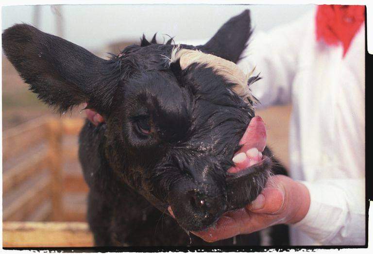
A fatal autosomal genetic defect that affects Black and Red Angus, Hereford, Simmental and Holstein • Calves are borne 10-30 days early • They show head abnormalities such as brachygnathia inferior, impacted molars and protruding tongue • The long bones of the affected animals are “shorter” • Bones are very fragile and can be easily broken • Caused by a deletion in SLC4A2 gene on chromosome 4
4.Hypotrichosis (Hairless Calf)
There is complete or partial loss of hair but it will grow like short curly coat hair • Calf has black diluted charcoal or chocolate colored hair • Affected animal is prone to environmental stress and skin infections • Affected gene is PMEL 17 (BTA5) encoding the premelanosome protein. • Defect is due to a mutation ( 3bp is deleted, CTT in exon 1) • Described in Angus, Ayrshire, Brangus, Holstein Friesian, Hereford, PolledHereford, Guernsey, Gelbvieh, Jersey, Normandy Maine, Charolais, and Simmental crosses. • Most have autosomal recessive and/or sex-linked mode of inheritance • 13 types is described in cattle
5.Hypotrichosis in other Livestock
Breed affected is the Polled-Dorset • Wool is of poor quality • HR gene is affected (1313C/T leads to 438Gln/Stop) • Hypotrichosis in goats is associated with goiter • In swine, 2 forms are known Mexican Hairless and German -associated with goiter and death of homozygote
6.Atresia Ani/Atresia Coli
This is the most common congenital defect of the lower GIT The affected animal is observed to have absence of anal opening This results when the dorsal membrane separating the rectum and anus fail to rupture In female lambs or kids, the terminal portion of the rectum may open into the vagina
7.Inguinal Hernia/Scrotal Hernia
The viscera pass does the inguinal canal and may lie in the cavity of the tunica vaginalis In scrotal hernia, there may be testicular degeneration
Common in male pigs, • In female pigs, genital development is arrested (animal becomes sterile) • In stallions, it is characterized by signs of constant and severe abdominal pain • In cattle, this defect is not common • Surgical correction is done to preserve the breeding potential of the animal but is not always successful
8.Albinism
Result of recent studies shows that it is an autosomal recessive disease • A single base substitution (G1431A), leads to conversion of tryptophan into a stop codon • This produces inactive or abnormal protein • The animal is characterised with white coat, non-pigmented skin and eyes, and pink mucosa • Non-pigmentation in the iris and retina leads to photophobia • True albinism is associated with pink or pale irises with visual defects and increased solar radiation-induced neoplasm of the skin • It is noted in Icelandic sheep, Guernsey, Austrian Murboden, Shorthorn, Brown Swiss and Charolais cattle.
9.Epitheliogenesis Imperfecta (Aplasia Cutis)
Lethal and there is lack of skin on the distal parts of the limbs, deformed ear due to auricular epithelial defect, defects in the integument of the muzzle • Related to the defective metabolism of fibroblasts impairing the nutrition of the epithelium • Seen in cattle (autosomal recessive trait), horses, swine and sheep
10.Arachnomelia
facial deformities characterised by short lower jaw and concave rounding of the dorsal profile of the maxilla is observed in affected animal • deformities in the distal part of the hind leg (bilateral hyperextesion of the fetlocks) • Defect is due to SUOX gene (BTA5) encoding molybdehemoprotein sulphite oxidase • Brown cattle – single base insertion (c.363-364insG) in exon 4 leading to premature stop • Simmental cattle – single base deletion (c.1224-1225delC)
11.Complex Vertebral Malformation (CVM)
A lethal genetic defect in a single recessive gene • Causes fetal resorption, abortion or stillbirth, have reduced fertility manifested as poor conception rate • Skeletal malformations such as shortened neck and thorax, deformed carpal and metacarpal joints, distortion, twisting and hypoplasia of the tail • The affected calf posses this mutation in both alleles, indicating autosomal recessive disorder • It is observed in Holstein breed of cattle This is due to single base substitution in SLC35A3 gene • Thee is missense (valine to phenylalanine) in genes SLC35A3 (Solute Carrier 35 Member 3) coding for uridinediphosphateN-acetylglucosamine transporter. • There is abnormal nucleotide-sugar transport into the Golgi apparatus, thus disrupting the normal protein glycosilation • Prevalence: – Turkey: 3.4% Denmark: 31% Poland: 24.8% – Japan: 32.5% Sweden: 23% Germany: 13.2%
12.Weaver Syndrome (Bovine Progressive Degenerative Myelopathy)
There is functional disturbance or pathological change in the spinal cord • Most commonly observed in brown Swiss cattle • Mutations in EZH2 (Histone-lysine N-Methyltransferase) • The animal have an odd weaving gait and this is due to weakness and lack of coordination in all limbs. • Clinical symptoms starts to appear at 6 months and becomes progressively worse until the animal dies
13.Spinal Dysmielination
Affects American Brown Swiss cattle • The SPAST gene (BTA11), encoding the spastin protein has a missense mutation c.560G>A (p.Arg560Glu) • A congenital neurodegenerative disease in cattle • Recumbency and spastic extension of the limbs • There is variable degrees of denervation atrophy in the skeletal musculature
14.Bovine Leukocyte Adhesion Deficiency (BLAD)
An immunological disorder caused by substitution of A to G at nucleotide 383 in the CD18gene (ITGB2) • This leads to replacement of aspartic acid with glycine at position 1228 in the glycogen protein (D128G) • The affected animal is observed to have frequent bacterial infection, delayed wound healing and stunted growth • Severe and recurrent mucosal infection such as pneumonia • The reproductive performance and milk production is poor • Most common among Holstein cattle Turkey: 4% Brazil: 2.8% Japan: 4% USA: 4% Poland: 3% Iran: 3.3%
15.Congenital Erythropoietic Porphyria
It is a hereditary enzyme deficiency in the haeme (essential part of hemoglobin) biosynthesis • The affected gene is the UROD gene (uroporphynogen decarboxylase) which encodes the enzyme uroporphyrinogen III synthase • There is photosensitization which causes subepidermal blistering and dermal necrosis • Affected animal have varying degrees of reddish-brown discoloration of bones, teeth and urine
16.Hereditary Zinc Deficiency (HZD
This defect is due to single substitution in gene SLC39A4, wherein there is impaired intestinal zinc absorption • It is characterised by paraketosis and dermatitis and it occurs in areas/regions that are subjected to abrasion • Lesions could be found around the mouth, eyes, base of the ear, joints, and lower parts of the thorax, abdomen and limbs.
17.Citrullinemia
Fatal hereditary metabolic defect of Holstein-Friesian calves (mainly in Australian and New Zealand) • This defect involves substitution of C-T in exon 5 of agrininosuccinate synthetase (ASS), which converts CGA codon (Arginine) to TGA (translational termination codon) • Clinical signs include ammonemia (increased circulatory ammonia) and related neurological signs • Calves may also present ataxia, aimless wondering, blindness, head pressing, convulsion and death • Frequency in US Holstein (0.3%) and Chinese Holstein (0.16%) • Case in Australia: due to importation of semen from US
18.Factor XI Deficiency (FXID)
Mutation in the F11 (Blood Coagulation Factor 11) caused the defect • There is 76bp insertion in the exon 12 in bovine chromosome 27 • FXID results to prolonged bleeding (after birth, dehorning, castration, etc.) • Affected cows frequently have pink-colored colostrum • It also reduced reproduction performance and animals affected are more susceptible to diseases (pneumonia, mastitis and metritis) • Affected animals have higher morbidity and mortality • This defect have significant economic impact in the dairy industry Turkey: 1.8%
19.X-linked Anhydrotic Ectodermal Dysplasia
An X-linked recessive disease • It is caused by 19 bp deletion in EDA, which encodes Ectodysplasia A, a protein involved in the formation of hair follicles and tooth bud • Clinical findings include hairlessness, tooth abnormalities, and reduced sweat glands • Only males present full form, whereas heterozygous females (carriers) are asymptomatic or show light symptoms (hypotrichosis and reduced number of teeth)
20.Parakeratosis (Edema Disease, Lethal trait A46)
An inherited defect caused by (a) Simple Autosomal lethal factor (b) Chromosomal anomaly • Associated with poor intestinal uptake of zinc therefore develops conjunctivitis, diarrhea and increase susceptibility to infection • Calf is normal at birth but develops paraketosis at 5 weeks old and evetually dies • Head and neck: thickened, with scales, cracks and fissures • Eye area: abraded
Compiled  & Shared by- Team, LITD (Livestock Institute of Training & Development)


Image-Courtesy-Google


Reference-On Request.
TO BE CONTINUED SUBSEQUENTLY IN NEXT POST


