


COMMON POULTRY DISEASES
Dr. Rashmi,* Assistant Professor, Department of Veterinary Pathology
Dr. Karishma Meena, P.G. scholar, Department of Veterinary Medicine
College of Veterinary and Animal Science, Navania, Udaipur
RAJUVAS, Bikaner
Corresponding author*:rashmi01meena31@gmail.com
INTRODUCTION
Poultry farming plays a significant role in global food production, but it is vulnerable to a wide range of diseases that can adversely affect the health of birds and the economic stability of the industry. Poultry diseases, both viral and bacterial, can lead to high mortality rates, decreased productivity, and increased veterinary and management costs. This review examines the most common poultry diseases, including Avian Influenza, Newcastle Disease, Marek’s Disease, Coccidiosis, Fowl Pox, and Infectious Bronchitis, providing an overview of their symptoms, lesions, diagnostic methods, and treatment strategies.
Viral infections such as Avian Influenza and Newcastle Disease often result in sudden outbreaks, requiring rapid response and control measures, including culling and vaccination. Bacterial diseases like Coccidiosis and Fowl Pox, on the other hand, often cause chronic symptoms and can be mitigated with proper management practices, hygiene, and antimicrobial treatments. The importance of timely diagnosis using modern techniques such as PCR testing and virus isolation is emphasized for effective disease management.
- Avian Influenza (Bird Flu)
Etiology:
Avian influenza or Bird flu is caused by influenza A viruses, with subtypes like H5N1 and H7N9.
Symptoms:
- Sudden death in some cases.
- Swelling of the head, neck, and eyes.
- Coughing, sneezing, and nasal discharge.
- Decreased egg production and soft-shelled eggs.
- Lethargy and a decrease in feed and water intake.
Lesions:
- Swelling and hemorrhage in various organs such as the heart, lungs, and liver.
- Cyanosis (blue discoloration) of comb and wattle.
- Lesions in the digestive tract, such as hemorrhage and necrosis.
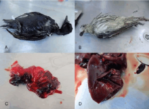
Fig:1. Post mortem lesions showing (A&B): dead birds infected with avian influenza (C) Congestive lung (D) Necrosis in the liver.
Diagnosis:
- PCR testingto detect the virus.
- Serologyto test for antibodies.
- Virus isolation in eggs or cell cultures.
Treatment:
- There is no specific treatment for avian influenza, but antiviral drugs may be used in some cases.
- Supportive care, including maintaining hydration and nutrition, can help reduce mortality.
- Culling of infected birds to prevent spread.
- Newcastle Disease
Etiology: Newcastle disease in chickens is caused by avian paramyxovirus type 1.
Symptoms:
- Sudden death.
- Coughing, sneezing, and nasal discharge.
- Tremors or paralysis (especially in legs and wings).
- Swollen eyelids and neck.
Lesions:
- Hemorrhagic lesions in the digestive tract, especially the ceca.
- Lesions in the brain and nervous system (encephalitis).
- Edema and swelling of the head and neck.
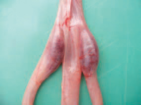
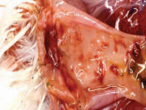
Fig:2 This photograph showing is the enlargement and haemorrhages of caecal tonsils and haemorrhagic cloacitis.
Diagnosis:
- PCR testingto confirm the presence of the virus.
- Isolation of the virus in cell cultures.
- Serology for antibodies.
Treatment:
- No specific antiviral treatment.
- Vaccination is the most effective way to prevent the disease.
- Supportive care, including providing proper hydration and nutrition.
- Marek’s Disease
Etiology:
Marek’s disease in chickens also known as fowl paralysis is caused by a highly contagious alpha herpesvirus.
Symptoms:
- Paralysis of wings, legs, or neck.
- Abnormal pupil shape or color in eyes ( “starry eyes”).
- Weight loss and reduced appetite.
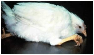
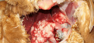
Figure 3: Leg paralysis of Marek’s Disease suspected chickens
Figure 4: This photograph showing tumor in spleen and liver
Lesions:
- Tumors in various organs, including the liver, lungs, heart, and spleen.
- Nerve damage and atrophy of nerves in the affected birds.
Diagnosis:
- Detection of the virus using PCR or tissue culture.
- Histopathology to identify characteristic tumors and nerve lesions.
Treatment:
- No cure, as Marek’s disease is caused by a virus.
- Vaccination at hatch is the best preventive measure.
- Culling of infected birds.
- Coccidiosis
Etiology:
Coccidiosis in poultry is caused by protozoan parasites from the genus Eimeria.
Symptoms:
- Bloody diarrhea.
- Lethargy and decreased feed intake.
- Weight loss and poor growth.
- Pale comb and wattle due to anemia.
Lesions:
- Lesions in the intestines, particularly in the ceca, showing areas of necrosis or hemorrhage.
- Swelling and inflammation in the affected areas of the intestine.
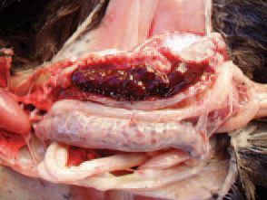
Fig: 5This photograph showing caeca are filled with fresh or clotted blood.
Diagnosis:
- Fecal examination for oocysts (eggs of the parasite).
- Postmortem examination of intestines to observe lesions.
Treatment:
- Anticoccidial drugs such as amprolium, sulfonamides, or ionophores.
- Maintaining good hygiene and sanitation to control the spread of the disease.
- Prophylactic use of anticoccidial drugs in feed.
- Fowl Pox
Etiology: Fowl pox in poultry is caused by pox virus (Avipox virus)
Symptoms:
- Lesions in the mouth, throat, and eyes (wet form) or on the skin (dry form).
- Swollen face and neck.
- Respiratory distress (for the wet form).
- Reduced egg production.
Lesions:
- Raised, wart-like lesions on the skin (dry form).
- White, caseous lesions in the mucous membranes of the mouth and respiratory tract (wet form).

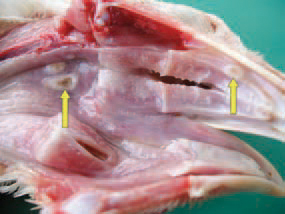
Fig:6 This photogtaph showing (A)wart-like lesions on the skin (dry form),(B) White, caseous lesions in the mucous membranes of the mouth and respiratory tract (wet form).
Diagnosis:
- Clinical signs and observation of lesions.
- Virus isolation or PCR testing.
Treatment:
- No specific treatment for the virus.
- Vaccination is the most effective preventive measure.
- Infected birds may recover with supportive care.
- Infectious Bronchitis
Etiology: Avian infectious bronchitis (IB) is caused by the Gamma coronavirus.
Symptoms:
- Coughing, sneezing, and nasal discharge.
- Decreased egg production.
- Watery or thin-shelled eggs.
- Swelling of the face and eyes.
Lesions:
- Inflammation in the respiratory tract (trachea and bronchi).
- Swollen kidneys.
- Lesions in the reproductive tract in laying hens.
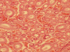
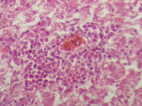
Fig:7 This photograph of Kidney showing (A) haemorrhages and (B) interstitial nephritis
Diagnosis:
- PCR testing to detect the virus.
- Serology and virus isolation.
Treatment:
- No specific antiviral treatment.
- Vaccination is essential to prevent the disease.
- Provide supportive care to reduce secondary infections.
- Salmonellosis
Etiology: Salmonella species (e.g., Salmonella enterica serovars like Salmonella enteritidis and Salmonella typhimurium).
Transmission:
- Direct contact with infected birds or contaminated feces.
- Contaminated feed, water, and equipment.
- Infected eggs (via the egg shell or the internal contents).
Symptoms:
- Diarrhea (sometimes with blood).
- Reduced egg production.
- Mortality (in severe cases).
- Swollen abdomen or organs (if it spreads to internal organs).
Lesions:
- In young birds usually include unabsorbed yolk sacs and classic gray nodules in the liver, spleen, lungs, heart, gizzard, and intestine.
- Firm, cheesy material in the ceca and raised plaques in the mucosa of the lower intestine are sometimes present.
- Occasionally, synovitis is prominent.

Fig:8 This photograph showing Multifocal granulomatous hepatitis in liver and nodular myocarditis
Diagnosis:
- Bacterial culture from feces or internal organs.
- PCR (Polymerase Chain Reaction) for detecting Salmonella DNA.
Prevention & Control:
- Good biosecurity measures.
- Maintaining proper hygiene (cleaning and disinfecting).
- Vaccination (in some cases, for certain strains like Salmonella enteritidis).
- Strict control of feeding practices and water sources to prevent contamination.
- Rodent and insect control to prevent spread.
- Colibacillosis (E. coli Infections)
Etiology: Escherichia coli (E. coli) bacteria
Transmission:
- Direct contact with infected birds or contaminated environments.
- Contaminated feed or water.
Symptoms:
- Respiratory signs (e.g., coughing, nasal discharge).
- Swollen abdomen.
- Diarrhea or watery stools.
- Sudden death in severe cases (e.g., septicemia).
- Reduced egg production in layers.
- Lameness (due to joint infections).
Lesions:
- Enlarged, hyperemic liver and spleen with increased fluid in body cavities.
- Subacute fibrinopurulent airsacculitis, pericarditis, perihepatitis, and lymphocytic depletion of the bursa and thymus.
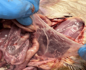
Fig:9 This photograph showing (fibrinopurulent airsacculitis) air sac is slightly more opaque than normal and contains some serous exudate.
Diagnosis:
- Isolation and identification of colifrom infected tissues or feces.
- Serotyping to identify specific strains.
Prevention & Control:
- Improved sanitation and biosecurity practices.
- Proper ventilation to reduce respiratory stress.
- Vaccination in certain cases (e.g., in layers).
- Probiotics or competitive exclusion strategies to support gut health.
- Antibiotic treatment under veterinary supervision.
- Mycoplasmosis
Etiology: Mycoplasma gallisepticum a bacterium without a cell wall.
Transmission:
- Direct contact between infected and susceptible birds.
- Airborne transmission (especially in densely populated poultry houses).
- Vertical transmission (from infected hens to offspring).
Symptoms:
- Respiratory distress (e.g., coughing, nasal discharge).
- Conjunctivitis (inflammation of the eyes).
- Swelling of the head and neck (in severe cases).
- Poor growth and reduced feed conversion in broilers.
- Reduced egg production and poor hatchability in layers.
- Mortality, especially in young birds.
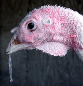
Fig: 10This photograph showing clear mucoid exudate, dried nasal exudate, and a swollen infraorbital sinus.
Lesions:
- Severe mucopurulent sinusitis and varying degrees of tracheitis and airsacculitis. Microscopically, mucous membranes are thickened, hyperplastic, necrotic, and infiltrated with inflammatory cells. The mucosal lamina propria contains focal areas of lymphoid hypoplasia and germinal center formations.
Diagnosis:
- PCR (Polymerase Chain Reaction) for detecting Mycoplasma DNA.
- Serological tests (e.g., ELISA or complement fixation test).
- Isolation and culture of the bacteria.
Prevention & Control:
- Strict biosecurity to prevent the introduction of infected birds.
- Segregation and isolation of infected flocks.
- Use of vaccines (particularly for layers) in areas where the disease is common.
- Antibiotic treatment (though Mycoplasma is generally resistant to many antibiotics).
- Avoid overcrowding to reduce stress, which can exacerbate the disease.
- Avian Cholera (Pasteurellosis)
Etiology: Pasteurella multocida, a highly pathogenic bacterium.
Transmission:
- Direct contact with infected birds.
- Contaminated water, feed, or equipment.
- Wild birds (especially ducks and geese) can be reservoirs and transmitters.
Symptoms:
- Sudden death (especially in acute cases).
- Swollen liver, spleen, and other organs.
- Diarrhea (with a greenish or watery appearance).
- Nasal discharge and respiratory distress (in chronic cases).
- Reduced egg production.
- Swollen joints and lameness in some cases.

Fig: 11 This photograph showing swelling in joints of adult birds
Lesions:
- Blue coloration of wattles, swollen wattles and face.
- Yellow-brown pus accumulated in a swollen wattle Pus (whitish to yellow) accumulated in a hock joint.
- Pinpoint bleeding in the muscles of heart.
Diagnosis:
- Bacterial culture and identification.
- PCR for detecting multocidaDNA.
- Postmortem examination revealing characteristic lesions (e.g., swollen liver).
Prevention & Control:
- Vaccination (available for specific strains of multocida).
- Strict hygiene and sanitation to prevent environmental contamination.
- Control access to wild birds.
- Use of antibiotics in the early stages of infection (under veterinary guidance).
- Fowl Typhoid
Etiology: Salmonella Gallinarum, a specific species of Salmonella.
Transmission:
- Fecal-oral route (via contaminated water or food).
- Direct contact with infected birds.
Symptoms:
- High fever.
- Loss of appetite.
- Diarrhea (often with a foul odor).
- Lethargy and weakness.
- Reduced egg production.
- Sudden death in severe cases.
Lesions:
- Swollen, friable, and often bile-stained liver, with or without necrotic foci
- Enlarged spleen and kidneys
- Anemia
- Enteritis
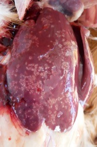
Fig:12 This photograph showing Enlarged liver with multiple white, necrotic lesions.
Diagnosis:
- Bacterial culture from feces or internal organs (e.g., liver, spleen).
- Serological tests (for antibody detection).
Prevention & Control:
- Vaccination of breeder flocks.
- Strict sanitation and good biosecurity practices.
- Rodent and insect control to limit spread.
- Quarantine and testing of incoming birds.
General Treatment Guidelines:
- Vaccination: Vaccination is one of the most effective methods to prevent many poultry diseases, including Newcastle disease, Marek’s disease, and avian influenza.
- Hygiene and Biosecurity: Regular cleaning and disinfection of equipment, housing, and areas where poultry are raised can significantly reduce the spread of diseases.
- Antibiotics/Antiviral Drugs: These may be used for secondary infections, but they are generally not effective against viral diseases like avian influenza or Marek’s disease.
- Culling: In severe cases where diseases cannot be controlled or are highly contagious, culling infected birds may be necessary to prevent the spread.