


UNDERLYING CAUSES OF AVIAN MYCOPLASMOSIS SYMPTOMS AND PREVENTIVE MEASURES
***
Dr. Rambabu.D, MVSc, Ph.D, MBA.
Associate Professor
Dept. of Poultry Science, College of Veterinary Science, Korutla
PV Narsimha Rao Telangana Veterinary University
Jagtial dist – 505 326. Telangana State.
***
Introduction
Chickens and turkeys are most commonly affected by the infectious respiratory disease known as avian mycoplasmosis. In the poultry business, it can result in large financial losses and is caused by different species of Mycoplasma. Both Mycoplasma synoviae (MS) and Mycoplasma gallisepticum (MG) are the two main Mycoplasma species that cause mycoplasmosis. Many illnesses of the respiratory system and the body in birds are caused by these bacteria. The primary feature that sets these bacteria apart is their absence of a cell wall. They are the most basic prokaryotic microbes capable of self-replication. Egg production usually decreases during the initial infection, recovers, and is maintained at a lower level in chickens infected with MG, the most common cause of mycoplasmosis in poultry. Infected chickens exhibit a wide range of symptoms, including rales, coughing, nasal discharge, conjunctivitis, reduced feed efficiency, and air sacculitis.
Infections with Mycoplasma gallisepticum in birds typically cause severe respiratory tract inflammation after a protracted incubation period. The illness frequently remains undetected in flocks and results in latent illnesses. A number of factors have been linked to the transmission and outbreak of disease, according to past research: high feed density, heat and cold stress, elevated ammonia levels, faeces accumulation, fouling of the chicken house, vast temperature swings, and sudden changes in the climate.
Factors influencing virulence
Numerous surface polypeptides and lipoproteins of the MG plasma membrane are known, and they may play a role in MG virulence factors including as motility, cytadhesins, surface antigen variation and nutrition acquisition.
Interaction between the host and pathogen
gallisepticum uses specific surface proteins to adhere to the ciliated cells in birds’ respiratory tracts. The attachment of MG to the host cell is crucial for successful colonisation and eventual pathogenesis. It infiltrates the host cells after it is attached. Mycoplasma gallisepticum species may penetrate target tissues and get beyond the physical defences of their hosts, such as ciliary activity and respiratory mucus, thanks to their gliding motility.
Multiplication and Colonisation: Since it appears to enhance persistent infection and host immune response evasion, emerging phenotypic variation—which is recognised as a role of several MG cytadhesins and putative cytadhesins—is thought to be a crucial component of virulence. The bacteria replicates inside the cells after it has invaded them.
The inflammatory reaction
When the immune system of the host detects the presence of the bacteria, an inflammatory response is triggered. To fight the infection, this entails the release of different immune cells and inflammatory mediators.
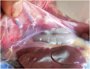
Lesions and Damage to Tissue
The host immune system, bacterial multiplication, and inflammation can all cause harm to the respiratory tissues. Lesions in the air sacs, bronchi, and trachea may be the result of this injury.
Transmission
Within a flock, M. gallisepticum can spread by respiratory secretions, faeces, infected tools or surroundings. Potential sources of MG infection include backyard flocks, multi age commercial layer flocks, and certain wild bird species. The conjunctiva and upper respiratory tract serve as entrance points for microorganisms in aerosols or droplets. Mycoplasma gallisepticum rarely survives outside the host for more than a few days, so the dynamics of MG infection primarily depend on clinically or sub clinically infected carrier birds. However, multiple studies have shown that MG can survive for up to several days on contaminated fomite materials (dust, feathers, etc.), which offers crucial information about the disease’s epidemiology. The ability of certain M. gallisepticum strains to form biofilms may allow them to withstand longer periods of time in the environment. Transovarian transmission is one way that MG can spread from an infected breeding flock to their offspring. According to several research, the acute phase of the disease is when vertical transmission of MG occurs at its maximum rates, peaking the level of MG in the respiratory tract, and then declining as the post-infection interval lengthens.
State of Carrier
Certain birds have the potential to harbour the bacteria M. gallisepticum without displaying obvious symptoms of disease. Other birds may contract the illness from these carriers.
Signs and symptoms
Mycoplasmosis-infected birds frequently display respiratory distress, which is typified by coughing, sneezing, nasal discharge and wheezing.
Decreased Production of Eggs: Mycoplasmosis in laying hens can result in a drop in both egg production and egg quality.
Swollen Eyes and Sinusitis: Infected birds may experience ocular discharge in addition to swelling and sinusitis.
Conjunctivitis
Conjunctival inflammation carried on by the infection may result in redness and discharge from the eyes.
Overall Weakness
Birds that are infected may become lethargic, lose their appetite, and become less active overall.
Diagnosis
A precise diagnosis is essential for the efficient treatment of mycoplasmosis. It is usually accomplished by combining laboratory testing, post-mortem investigations, and clinical indicators. Polymerase chain reaction (PCR), serology (blood testing), and bacterial isolation from afflicted tissues are some examples of these tests.
Prevention and Control
Bio security Operations
Take stringent bio security precautions to stop mycoplasmosis from entering and spreading. This entails restricting visitor access, keeping workers’ shoes and clothes separate, and routinely sanitising facilities and equipment.
Immunisation
Choose the best vaccination regimen for your poultry based on the available vaccines for both MG and MS.
Programmes for Cleanup
Better outcomes could be obtained by using the right chemical to effectively remove mycoplasmal infection before immunisation.
Reduce Stress
Because stress impairs immunity, birds are more prone to illness. By guaranteeing appropriate nourishment, ventilation, and living conditions, you may create a low-stress environment.
Monitoring
Keep an eye out for any indications of disease in your flock. Early diagnosis minimises the disease’s spread and enables timely treatments.