Foot and mouth disease in swine: Transmission, pathology, differential diagnosis and control measures
Das T1, Vidya Rani HB2, Das NK3, Sahoo M1 and S Mallick1
1ICAR-NIFMD, Bhubaneswar
2ICAR-IVRI, Izatnagar, Uttar Pradesh, India
3BVO, Bisoi, Mayurbhanj, FARD, Government of Odisha
Corresponding author E mail id: tarenisahoo@gmail.com
Foot and mouth disease is one of the highly contagious transboundary disease of ruminants and swine. Pigs generally get infected by oropharyngeal route after ingestion of FMDV contaminated feed or after direct contact with FMD affected animals or when placed in a heavily contaminated environment. Pigs are less susceptible to aerosol infection. Infected pigs produce 60 times more aerosol virus than the ruminants. Adult pigs usually suffer from chronic lameness and younger pigs die before exhibiting any clinical signs due to acute or hyper acute myocarditis. FMD in swine should be differentiated from other vesicular diseases like swine vesicular disease, vesicular stomatitis and vesicular exanthema. Elimination of source of infection, movement restriction, sanitary measures, zoning, isolation, conventional and emergency vaccination programs and import control are helpful measures for control and prevention of FMD.
Introduction
Foot and mouth disease is one of the highly contagious and the most serious transboundary viral disease of cloven-footed animals including ruminants and suids hindering livestock production causing severe global economic losses. Pig is the most common domestic animal raised worldwide and FMD affects all age groups of pigs. The economic losses due to FMD outbreak in Taiwan’s pig industry was estimated to be US$ 1.6 billion in 1997 (Yang et al., 1999). The financial losses in 180 piggery farms of the Republic Korea due to FMD outbreak in 2015/2015 was US$25.2 million (Yoon et al., 2018). FMD is caused by foot and mouth disease virus under Genus Aphtho virus under family Picornaviridae. It has seven serotypes. Serotype O is the most prevalent worldwide. Different isolates show different host range. Porcinophillic nature of virus is associated with shortened 3A NSP which is the characteristic of Cathay topotype (O/TAW/97) (Pacheco and Mason, 2010). Korean strain O/SKR/AS/2002 is highly virulent to pigs and limited in cattle but has intact 3A region (Oem et al., 2002).
FMD transmission in pigs
FMD pathogenesis vary in different species in several aspects. Pigs are less susceptible to aerosol infection. They require 600 times more than the exposure to aerosol virus required by bovine or ovine. Porcine nasopharynx is less permissive to FMDV as compared to oropharynx (Fukai et al., 2015; Stenfeldt et al., 2016). Pigs generally get infected by oropharyngeal route after ingestion of FMDV contaminated feed or after direct contact with FMD affected animals or when placed in a heavily contaminated environment (Kitching and Alexandersen, 2002). The onset and progression of disease was more rapid in pigs exposed to donor infected pigs during advance stage of the disease (Stenfeldt et al., 2016). FMDV remains viable and infectious in contaminated pig feed for a longer period of time (more than 37 days) compatible with transoceanic transmission (Stenfeldt et al., 2022). In an in -vivo study, it was demonstrated that intradermal route is more effective in transmitting FMDV to pigs than intravenous route (Quan et al., 2004). Also, on increasing the exposure intensity by increasing the numbers of pigs/ infected pigs housed together shortens the time for onset of clinical disease in pigs (Quan et al., 2004). Infected pigs produce 60 times more aerosol virus than the ruminants (Alexandersen et al., 2003) and also aerosol production varies with different FMDV strains. In an experimental study, it was demonstrated that less exposure time is required for strain A24 Cru than strain O1 Manisa and Asia1 Shamir. Also, the animal infected with A24 Cru exhibited highest level of virus shedding than strain O1 Manisa and Asia1 Shamir (Pacheco et al., 2012). More aerosol excretion in pigs is associated with development of clinical signs which fades away as antibody develops (Kitching and Alexandersen, 2002). Also, pigs are more capable of clearance FMDV infection in contrast to ruminants, therefore in pigs no subclinical carrier stage has been reported (Stenfeldt et al., 2016).
Pathogenesis of FMD in pigs
During the early natural infection, the suggested sites for virus replication in pigs are soft palate, tonsil and the floor of the pharynx and then spread of virus from pharyngeal region to the regional lymph nodes and then to stratified squamous epithelial cells/ Langerhans cells via blood infecting only few cells that result in major amplification of virus and higher viraemia and then infecting higher numbers of cells (Alexandersen et al., 2001; Murphy et al., 2010). The replication cycle is repeated several times (Alexandersen et al., 2001). The stratified squamous epithelium of skin, oral mucosa and pharynx are mostly responsible for virus replication (Alexandersen et al., 2001). The reticular epithelium of oropharyngeal tonsil particularly paraepiglottic tonsil is the primary site for early virus replication (6hpi). The reticular epithelium or MALT associated epithelium has discontinuous basement membrane and abundant transmigrating leucocytes (Stenfeldt et al., 2016). The virus antigen was demonstrated in the cytokeratin positive epithelial cells and CD172a expressing leucocytes of oropharyngeal tonsil (Stenfeldt et al., 2014).
Clinical signs and pathology of FMD in pigs
The incubation period of FMD in pigs varies from 24 hours to 14 days (Quan et al., 2004). Both feral pigs and domestic pigs are susceptible to FMD. But feral pigs are more tolerant to FMD than domestic pigs. In an experimental study, domestic pigs developed clinical signs within 24 hours of contact but feral pigs did not develop any clinical signs until 48 hours. Also, the vesicular lesions are difficult to detect due to dark and thick skin in feral pigs (Mohamed et al., 2011). However, they exhibited similar clinical signs and excreted similar amount of virus.
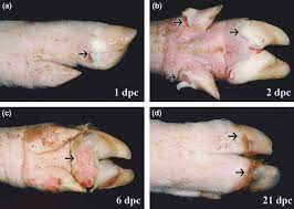
The clinical signs in pigs are fever, local signs of inflammation on areas of feet, blanching of coronary bands, lameness, unable to stand and walk, swollen heel bulbs, development of vesicular lesions in the coronary bands, heel bulbs, lower jaw, mouth, tongue and snout. The affected pigs are lethargic, anorexic and huddled together. Lesions on the coronary band are the most consistent findings (Kitching and Alexandersen, 2002). The peeling of skin or rupture of the vesicular lesion usually occurs within 24-48 hours resulting in red raw surface. Secondary bacterial infection may cause abscess formation that delays healing. Sometimes there are sloughing of claws (Mohmed et al., 2011). Adult pigs usually suffer from chronic lameness and younger pigs die before exhibiting any clinical signs due to acute or hyper acute myocarditis (Kitching and Alexandersen, 2002). Sows may abort. Histopathologically, there is ballooning degeneration, intercellular oedema and disruption of the stratum spinosum layer of the epidermis and occasionally stratum basale layer with infiltration of lymphocytes and plasma cells in the underlying dermis. But secondary bacterial infection may result in accumulation of neutrophils and bacterial colony (Mohmed et al., 2011). Necrosis of heart muscle cells with lymphohistocytic infiltration are detected both grossly and microscopically in the heart of young animals. In situ hybridization study recognized the highest signal for detection of positive and negative strand of FMDV RNA in the basal cell layer of both tongue and skin epithelium suggestive of early replication site for FMDV. ISH also demonstrated diffuse positive signal of positive strand FMDV RNA in the stratum spinosum layer (Durand et al., 2008).
Diagnosis of FMD in pigs
FMD can be diagnosed on the basis of clinical signs. FMD in swine should be differentiated from other vesicular diseases like swine vesicular disease, vesicular stomatitis and vesicular exanthema. Swine vesicular disease (SVD) is caused by Enterovirus genus under family Picornaviridae. It is transmitted through broken skin and mucosa, ingestion of contaminated meat, inhalation and contact with contaminated faeces and other excretions. It produces similar lesions like FMD but often mild and sub clinical in nature. Vesicular stomatitis (VS) is caused by Vesiculovirus under family Rhabdoviridae and it has two serotypes Indiana and New Jersey. It is mechanically transmitted by flies. Clinical signs are similar to FMD but it is less contagious than FMD. In VS, no lesions are observed in heart. Lameness and foot lesions are observed first in pigs affected with VSV. VS is suspected when cattle, horse and pigs are affected concurrently as horse is resilient to FMD. Sheep and goats are resistant to vesicular stomatitis. Vesicular exanthema (VESV) is caused by Vesivirus under family Calciviridae. It is transmitted by feeding of uncooked garbage and fish scraps. It is highly infectious disease but with less mortality. VESV is associated with vesicular disease like FMD, reproductive failure and mild encephalitis in pigs. Therefore, confirmatory laboratory diagnosis for FMD is essential.
There are many techniques for differential diagnosis of FMD from other vesicular diseases like RT-PCR system for differential diagnosis of different vesicular diseases of swine (Nunez et al., 1998), one step multiplex RT-PCR (Fernandez et al., 2008), m RT- qPCR for simultaneous detection of FMD, African and classical swine fever virus (ASFV& CSFV) from swine oral fluids (Grau et al., 2015) etc. Different nucleic acid detection techniques are available for detection of FMD RNA from different clinical samples like RT-PCR using porcine pedal vesicle, serum and saliva; RT-ii PCR using nasal, oral swabs and vesicular fluid from piglets; multiplex RT-q PCR assay for simultaneous detection of different serotypes; RT-PCR micro array using serum, nasal and oral swabs for differential diagnosis of FMDV, SVD, VESV, ASFV, CSFV, PCV2 and PRRSV etc.; RT-LAMP, multiplex RT-LAMP, real time RT-LAMP etc. from different clinical samples (Wong et al., 2020). Apart from molecular techniques, different serological methods are used for detection of FMD antibodies like virus neutralization test, indirect ELISA, sandwich ELISA, liquid phase blocking ELISA, solid phase competitive ELISA, microchip-based ELISA, NSP based ELISA using pig serum samples (Wong et al., 2020). Lateral flow dipstick based on recombinant ABC3 was developed for detection of anti-NSP antibody in FMD infected swine (Chen et al., 2009) and multiplex lateral flow strip was developed for simultaneous detection of seven FMD virus serotypes (Morioka et al., 2015). Swine oral fluid is a potential material for detection of FMDV genome by q RT-PCR (1-21dpi), for virus isolation (1-5dpi), for antigen detection by lateral flow immunochromatographic strip test (1-6 dpi) and by double antibody sandwich ELISA (2-3dpi), for detection of FMD specific mucosal IgA (14dpi) using isotype specific ELISA (Senthilkumaran et al., 2015).
Control and prevention
In uninfected pig herd, FMD can be controlled by vaccination of 12-14 weeks old pigs and repeated vaccination 2 weeks later. Vaccination of pregnant sows will provide passive immunity to younger piglets through colostrum. In presence of clinical disease, vaccination alone is not sufficient to provide protection. In FMD free countries, FMD control is carried out by slaughter of all affected pigs and in contact susceptible pigs, movement restriction and disinfection (Kitching and Alexandersen, 2002). For safe disposal of pig carcasses infected with FMD, composting is a best method to inactivate virus (Guan et al., 2010). FMD remains viable in contaminated pig feed products through 37 days and the infection risk varies with type of contaminated feed, the virus strain, the feeding condition, storage temperature etc. Pre-treatment of feed with feed additives like formaldehyde or lactic acid can prevent FMD infection (Stenfeldt et al., 2022). Elimination of source of infection through effective epidemiological surveillance, disturbing contact between infected and susceptible animals through movement restriction, sanitary measures, zoning, isolation etc., decreasing the numbers of susceptible animals via conventional and emergency vaccination programs in endemic areas and areas with recent FMD introduction and import control through quarantine in FMD free areas are helpful measures for control and prevention of FMD (Leo et al., 2012).
References
Alexandersen S, Oleksiewicz MB, Donaldson AI. (2001). The early pathogenesis of foot-and-mouth disease in pigs infected by contact: a quantitative time-course study using TaqMan RT–PCR. Journal of General Virology; 82(4):747-55.
Alexandersen S, Zhang Z, Donaldson AI, Garland AJ. (2003). The pathogenesis and diagnosis of foot-and-mouth disease. Journal of comparative pathology ;129(1):1-36.
Durand S, Murphy C, Zhang Z, Alexandersen S. (2008). Epithelial distribution and replication of foot-and-mouth disease virus RNA in infected pigs. Journal of comparative pathology; 139(2-3):86-96.
Fernández J, Agüero M, Romero L, Sánchez C, Belák S, Arias M, Sánchez-Vizcaíno JM. (2008). Rapid and differential diagnosis of foot-and-mouth disease, swine vesicular disease, and vesicular stomatitis by a new multiplex RT-PCR assay. Journal of virological methods ;147(2):301-11.
Fukai K, Yamada M, Morioka K, Ohashi S, Yoshida K, Kitano R, Yamazoe R, Kanno T. (2015). Dose-dependent responses of pigs infected with foot-and-mouth disease virus O/JPN/2010 by the intranasal and intraoral routes. Archives of virology; 160:129-39.
Grau FR, Schroeder ME, Mulhern EL, McIntosh MT, Bounpheng MA. (2015). Detection of African swine fever, classical swine fever, and foot-and-mouth disease viruses in swine oral fluids by multiplex reverse transcription real-time polymerase chain reaction. Journal of Veterinary Diagnostic Investigation ;27(2):140-9.
Guan J, Chan M, Grenier C, Brooks BW, Spencer JL, Kranendonk C, Copps J, Clavijo A. (2010). Degradation of foot-and-mouth disease virus during composting of infected pig carcasses. Canadian Journal of Veterinary Research;74(1):40-4.
Horak S, Killoran K, Leedom Larson KR. (2016). Vesicular exanthema of swine virus. Swine Health Information. Center and Center for Food Security and Public Health, 2016.
Kitching RP, Alexandersen S. (2002). Clinical variation in foot and mouth disease: pigs. Rev Sci Tech. 21(3):513-8.
León EA. (2012). Foot-and-mouth disease in pigs: current epidemiological situation and control methods. Transbound Emerg Dis. 59 Suppl 1:36-49.
Morioka K, Fukai K, Yoshida K, Kitano R, Yamazoe R, Yamada M, Nishi T, Kanno T. (2015). Development and evaluation of a rapid antigen detection and serotyping lateral flow antigen detection system for foot-and-mouth disease virus. PLoS One. 10(8): e0134931.
Murphy C, Bashiruddin JB, Quan M, Zhang Z, Alexandersen S. (2010). Foot‐and‐mouth disease viral loads in pigs in the early, acute stage of disease. Veterinary Record. 166(1):10-4.
Nishi T, Fukai K, Masujin K, Kawaguchi R, Ikezawa M, Yamada M, Nakajima N, Komeno T, Furuta Y, Sugihara H, Kurosaki C, Sakamoto K, Morioka K. (2022). Administration of the antiviral agent T-1105 fully protects pigs from foot-and-mouth disease infection. Antiviral Res. 208:105425.
Núñez JI, Blanco E, Hernández T, Gómez-Tejedor C, Martı́n MJ, Dopazo J, Sobrino F. (1998). A RT-PCR assay for the differential diagnosis of vesicular viral diseases of swine. Journal of virological methods. 72(2):227-35.
Oem JK, Yeh MT, McKenna TS, Hayes JR, Rieder E, Giuffre AC, Robida JM, Lee KN, Cho IS, Fang X, Joo YS. (2008). Pathogenic characteristics of the Korean 2002 isolate of foot-and-mouth disease virus serotype O in pigs and cattle. Journal of comparative pathology. 138(4):204-14.
Pacheco JM, Mason PW. (2010). Evaluation of infectivity and transmission of different Asian foot-and-mouth disease viruses in swine. Journal of veterinary science. 11(2):133-42.
Pacheco JM, Tucker M, Hartwig E, Bishop E, Arzt J, Rodriguez LL. (2012). Direct contact transmission of three different foot-and-mouth disease virus strains in swine demonstrates important strain-specific differences. The Veterinary Journal. 193(2):456-63.
Quan M, Murphy CM, Zhang Z, Alexandersen S. (2004). Determinants of early foot-and-mouth disease virus dynamics in pigs. Journal of comparative pathology. 131(4):294-307.
Senthilkumaran C, Yang M, Bittner H, Ambagala A, Lung O, Zimmerman J, Giménez-Lirola LG, Nfon C. (2017). Detection of genome, antigen, and antibodies in oral fluids from pigs infected with foot-and-mouth disease virus. Can J Vet Res. 81(2):82-90
Stenfeldt C, Bertram MR, Meek HC, Hartwig EJ, Smoliga GR, Niederwerder MC, Diel DG, Dee SA, Arzt J. (2022). The risk and mitigation of foot‐and‐mouth disease virus infection of pigs through consumption of contaminated feed. Transboundary and emerging diseases;69(1):72-87.
Stenfeldt C, Pacheco JM, Brito BP, Moreno-Torres KI, Branan MA, Delgado AH, Rodriguez LL, Arzt J. (2016). Transmission of foot-and-mouth disease virus during the incubation period in pigs. Frontiers in Veterinary Science.3:105.
Stenfeldt C, Pacheco JM, Rodriguez LL, Arzt J (2014) Early Events in the Pathogenesis of Foot-and-Mouth Disease in Pigs; Identification of Oropharyngeal Tonsils as Sites of Primary and Sustained Viral Replication. PLOS ONE 9(9): e106859.
Tsu-Han CH, Chu-Hsiang PA, Ming-Hwa JO, Hsiu-Min LI, Yu-Liang HU, Kuang-Pin HS, Parn-Hwa CH, Fan LE. (2009). Virology: Development of a chromatographic strip assay for detection of porcine antibodies to 3ABC non-structural protein of foot-and-mouth disease virus serotype O. The Journal of Veterinary Medical Science. 71(6):703-8.
Yoon H, Jeong W, Han JH, Choi J, Kang YM, Kim YS, Park HS, Carpenter TE. (2018). Financial Impact of Foot-and-mouth disease outbreaks on pig farms in the Republic of Korea, 2014/2015. Preventive veterinary medicine. 149:140-2.


