


HORMONAL DERMATOSIS IN CANINE
Aditya Kumar1, Satish Kumar2, Simran jeet Singh3* and Aditi Kuriyal4
¹PhD Scholar, Department of Veterinary Medicine
2Assistant Professor, Veterinary Clinical Complex
3PG Scholar, Department of Veterinary Medicine
4UG Scholar ,College of Veterinary and Animal Sciences ,G.B. Pant University of Agriculture and Technology, Pantnagar, Uttarakhand, India- 263145
Corresponding author mail- simranjeets140@gmail.com
Introduction:
A set of skin diseases in dogs known as hormonal dermatosis are impacted by hormone imbalances in the dog’s body. These ailments can lead to a variety of skin and coat issues, which can be upsetting for both the dog and the owner.
Etio-pathogenesis:
There are several types of hormonal dermatosis in canines, each associated with specific hormonal imbalances. Some common types include:
- a) Hypothyroid Dermatosis: Caused by hypothyroidism, a deficiency of thyroid hormones. It leads to xeroderma, alopecia, and increased susceptibility to skin infections.
Thyroxine, often known as T4, is a hormone generated by the thyroid gland that plays a critical role in regulating metabolism and many physiological processes throughout the body, including the skin. When the thyroid gland is underactive and doesn’t produce enough thyroxine (hypothyroidism), it can lead to several complications, including skin problems as Thyroxin is a requisite for the normal growth and maintenance of hair along with maintaining skin’s natural moisture balance. Thyroxine basically initiates hair growth.
Hair follicles may become less active in hypothyroidism, which can cause hair loss, coat thinning, changes in the texture of the hair that is still present and conditions like xerosis cutis. Thyroxine is also involved in regulating the production of melanin, which gives color to the skin and hair. Hypothyroidism can cause changes in skin pigmentation, leading to lighter or darker patches. Insufficient levels can result in thin, fragile skin that is more prone to injuries and infections and healing tendencies prone to diminish.
b) Hyperadrenocorticism (Cushing’s Disease) Dermatosis: Resulting from excessive production of cortisol due to adrenal gland dysfunction. Alopecia, dermatoporosis, dryness, increased susceptibility to infections, and the development of blackheads or comedones. The following dog breeds have a higher incidence of Cushing’s Syndrome: Beagles, Boston Terriers, Yorkies, Small Dachshunds, Poodles German shepherd, Large Poodles, Dachshunds.
c) Sex Hormone Dermatosis: Imbalances in sex hormones (estrogen and testosterone) can cause to alopecia (hair loss), changes in coat texture and color, and skin-related disorders. In female dogs, fluctuations in estrogen levels throughout the estrous cycle or during pregnancy can contribute to changes in the skin, coat, and even the formation of mammary tumors. Testosterone, on the other hand, can alter sebum (skin oil) production and the number of sebaceous glands in the skin. Increased testosterone levels might lead to overactive sebaceous glands, causing oily skin, acne-like conditions, and increased susceptibility to bacterial and fungal infections.
d) Diabetes Mellitus-Associated Dermatosis: Poorly controlled diabetes leads to elevated blood sugar levels which further leads to dehydration causing the skin to lose its normal moisture content and increase its dryness and appear flakey, and due to dryness dog might experience increased itching and scratching. Diabetes also results in poor wound healing and impaired immunity hence dogs with Diabetes are more prone to bacterial, yeast and fungal infections. (J Am Vet Med Assoc 2001:219: 203–208)
e) Growth Hormone-Associated Dermatosis: Growth hormone levels that are out of balance can alter the dog’s coat, frequently giving it a rough or “wooly” appearance. Each type of hormonal dermatosis has unique symptoms that may call for a particular approach to diagnosis and therapy. To effectively treat and manage these cases, one must correctly identify the underlying hormonal imbalance.
Clinical Signs & Symptoms:
Different cutaneous indications of endocrine illness can appear in dogs. Some indications may not be condition-specific; for instance, demodicosis, secondary skin infections (bacteria and yeast), and truncal alopecia with comedones (blackheads) can all be brought on by both Cushing’s disease and hypothyroidism. As an example, rat tail in dogs can be linked with hypothyroidism and Acanthosis nigricans with Diabetes mellitus. In addition, in these patients, alopecia is primary and not secondary to pruritus.
Common symptoms of hormonal dermatosis in canines include:
- Folliculitis
- Gradual Hair loss or thinning of fur coat (black skin disease)
- Erythema and swelling
- Dry, flaky, or scaly skin (Xeroderma)
- Calcinosis cutis in case of Cushing’s Syndrome
- Hyperpigmentation, “Acanthosis Nigericans,” characterized by dark, thickened, and velvety skin patches, often found in the armpits, groin, and neck. This can also occur in the folds of the skin.
- Recurrent skin infections
- Changes in coat color or texture (reduced lushness)
Itching and scratching which can exacerbate secondary skin infections and Secondary skin lesions like papules, pustules, and crusts.
Calcinosis cutis in a dog with Cushing’s Syndrome
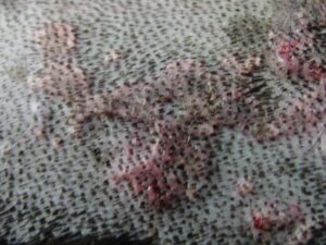
Image Source: Caroldermoid
Hyperpigmentation
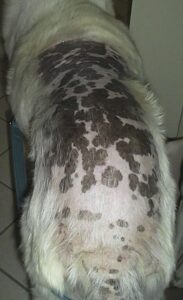
Image Source: Dr. Carl Palazzolo, DVM
Diagnosis: Diagnosing hormonal dermatosis in canines requires a comprehensive approach involving several steps
Medical history: This covers past skin conditions, alterations in behaviour, the environment, diet, and medication usage. If there is a history of persistent skin issues that have not responded to treatment, hormonal dermatosis may be suspected.
Physical Examination: The dog’s skin and coat should be thoroughly examined physically to look for any signs of hormonal imbalance induced Dermatosis, such as hair loss, thinning, redness, inflammation, scaling, crusting, or lesions, particularly in areas like the neck, flanks, tail base, and perineum.
Clinical Signs: Hormonal dermatosis can present in different ways depending on the specific hormonal imbalance. For example:
Hypothyroidism: Hair loss, lethargy, weight gain, and thickened skin.
Hyperadrenocorticism (Cushing’s disease): Bilateral hair loss, thin skin, increased appetite, and excessive thirst.
Hyperestrogenism: Swollen vulva, hair loss, and changes in behavior.
Blood Tests: To assess hormone levels, blood tests are essential. A comprehensive thyroid panel including T3, T4, and thyroid-stimulating hormone (TSH) is advised for hypothyroidism. Testing for cortisol levels, such as the low-dose dexamethasone suppression test, may be necessary in cases of Cushing’s illness. Depending on the symptoms, further hormone panels can be required.
Urinalysis: A urinalysis can help assess kidney function and identify any abnormal hormone levels that might be contributing to the skin issues.
Skin Scraping: Perform skin scrapings and combing of hair coat (flea combing) to rule out the presence of external parasites like mites, which can sometimes mimic hormonal dermatosis symptoms.
Skin Biopsy: In cases where other tests are inconclusive, a skin biopsy might be necessary. A biopsy can help identify specific changes in the skin layers that are characteristic of hormonal dermatosis.
Endocrine Testing: Depending on the suspected hormonal issue, additional endocrine testing might be needed. For instance, a low-dose dexamethasone suppression test can aid in diagnosing Cushing’s disease.
Treatment:
Treatment will be determined by the precise hormonal imbalance found when a diagnosis has been made. This could entail addressing secondary infections or skin conditions that emerged as a result of the hormonal imbalances, hormone replacement therapy, drugs to modulate hormone levels, dietary alterations, and more. Typical methods include:
Hypothyroidism Treatment: If hypothyroidism is diagnosed, oral thyroid hormone supplementation is prescribed to restore normal hormone levels.
Dosage & Route: Levothyroxine @ 0.02 mg/kg PO BID daily for 4-8 week
Cushing’s Disease Management: Management of Cushing’s disease may involve medication, surgery, or radiation therapy, depending on the underlying cause.
Medications like trilostane @ 2.2-6.6 mg/kg PO OD can help manage the condition by suppressing excess hormone production. Surgery might be considered in certain cases to remove adrenal tumors.
Sex Hormone Imbalance Correction: In cases of sex hormone imbalances, spaying or neutering may be recommended to regulate hormone levels.
Symptomatic treatment for skin lesions, Secondary bacterial Dermatitis, mycotic pruritus, and parasitic allergy due to hormonal Dermatosis are treated treated with antibacterial therapy, acaricides, and antifungals respectively after correcting the primary cause of ailment.
Multivitamin supplements along with omega fatty acids are administered for better recovery.
Dried up lesions are to be corrected by Keratolytic solutions and topical lotions and shampoos.
Preventive Measures:
While hormonal dermatosis cannot always be prevented, the risk can be reduced by ensuring general canine health. This entails supplying balanced food, consistent exercise, and standard care. Hormonal imbalances can also be prevented or managed by early detection and therapy.
References –
- Porter, R. S., & Kaplan, J. L. (Eds.). (2011). The Merck manual of diagnosis and therapy (19th ed.)
- Rory Breathnach, MVB, PhD, MRCVS (2008), Unusual Endocrine Dermatoses in the Dog
- Hill, P.B., Lo, A., Eden, C.A.N., Huntley, S., Morey, V., Ramsey, S., Richardson, C., Smith, D.J., Sutton, C., Taylor, M.D., Thorpe, E., Tidmarsh, R. and Williams, V. (2006). Survey of the prevalence, diagnosis, and treatment of dermatological conditions in small animals in general practice. Vet. Rec. 158:533-539.
- Scott, D. W., Miller, W. H. and Griffin, C. E. (2001). Small Animal Dermatology. (6th edn.), W. B. Saunders, Philadelphia.