


Important Bacterial Diseases in Poultry: Challenges and Solutions
Rahul. G. Kadam1*, Demian C. Johnson2, Anupam Brahma3, Saurabh Karunamay4, Privanka S. Narwade5
1*Assistant Professor, Unit of Teaching Veterinary Clinical complex (Veterinary Pathology), FVAS, I.Ag.Sc., RGSC, Banaras Hindu University, Mirzapur, U.P.:- 231307.
2Assistant Professor, Department of Veterinary Extension, FVAS, I.Ag.Sc., RGSC, Banaras Hindu University, Mirzapur, U.P.:- 231307.
3Assistant Professor, Department of Veterinary Parasitology, FVAS, I.Ag.Sc., RGSC, Banaras Hindu University, Mirzapur, U.P.:- 231307.
4Assistant Professor, Department of Livestock Products Technology, FVAS, I.Ag.Sc., RGSC, Banaras Hindu University, Mirzapur, U.P.:- 231307.
5PhDStudent, Department of Veterinary Gynecology & Obstetrics, FVAS, I.Ag.Sc., RGSC, Banaras Hindu University, Mirzapur, U.P.:- 231307.
*Corresponding author- rahulkadam@bhu.ac.in
Abstract
Poultry farming plays a crucial role in meeting the global demand for protein-rich food. However, bacterial,viral, fungal etc. diseases pose significant challenges to the poultry industry, leading to economic losses and public health concerns.Half of mortality in broiler breeders and commercial layers which is because of the non–outbreak-related bacterial diseases.It causes approximately 50% mortality during first week of a broiler’s life and in some instance,cause server outbreak eradicating almost all flocks. It causes production losses in subclinical infections besides increased mortality, also these forms as secondary invaders in most of the viral infections. The bacterial diseases of poultry are like; colibacillosis, fowl typhoid, paratyphoid infections (Avian salmonellosis), fowl Spirochaetosis, avian Pasteurellosis, Pullorumdiseases (Bacillary white diarrhoea of chicks),gangrenous dermatitis, avian Mycoplasmosis, necrotic enteritis,infectious coryza, chronic respiratory diseases and clostridial infection in poultry,out of thesewe explorefirst three important diseaseaffecting poultry, their causes, impacts, and strategies for prevention and control.
Colibacillosis
Bacteria of the species Escherichia coli are normal inhabitants of the digestive tract of mammals and birds and most strains are nonpathogenic. Colibacillosis refers to any localized or systemic infection caused by avian pathogenic Escherichia coli (APEC). The water is contaminated by the faces of birds. The produce different syndromes which includecolisepticemia, hemorrhagic septicemia, coligranuloma (Hjarre’s disease), airsacculitis (chronic respiratory disease, CRD), swollen‐head syndrome, venereal colibacillosis, coliform cellulitis (inflammatory or infectious process), peritonitis, salpingitis, orchitis, osteomyelitis/synovitis (including turkey osteomyelitis complex), panophthalmitis, omphalitis/yolk sac infection, and enteritis.
Members of the genus Escherichia are Gram-negative, flagellated rods (2x 0.6 microns). Grow on blood agar, ferment glucose, mannitol and lactose. E. coli serotypes aresomatic antigen O, flagellar antigen H and capsular antigen K. O antigen liberate endotoxin on autolysis of somatic cell comprised of a polysaccharide phospholipid complex boiling resistant protein factor. On the surface of the cell K antigen is present which is responsible for the virulence. K antigen further divided in to L and B forms based on its heat stability. Flagellar H antigen have no correlation with pathogenesis.
COLI-SEPTICAEMIA
Coli-Septicaemia exhibit seriousform of colibacillosis and with development and expansion of the broiler industries largenumbers of birds were kept intensively at high stocking rates in poorly ventilated
houses which result in eminence of severe form of diseases. This is acute infectious diseases usually affecting birds of 4 and 12 weeks of age. Birds shows clinical signs like ruffled feathers, laboured rapid berating and gasping with characteristic sound. The disease results in to approximately 5% mortality and 50% morbidity.
Pathogenesis
E.coli are always found in the digestive tracts of poultry and in particularly large numbers in the lower part of the small intestine and caecum and also because of E. coli containing dust inhalation,it found in throat and upper trachea. E. coli respiratory tractinfection along with other respiratory pathogens produce characteristic illness.Sometimes as a primary pathogenwhen resistance is lowered by environmental stress and poor air quality (especially dustor high ammonia levels) frequently causing colisepticaemia. sometimes also infect skin wounds or lesions leading to significant subcutaneous infections.
Postmortem findings
The distinguishing lesions like air sacculitis, peritonitis, perihepatitis and pericarditis are found regularly in postmortem. White caseous deposits found in air sacs which are thickened and opaque. Fibrinous pericarditis which is a characteristic feature of these diseases. There is thickening of pericardial sac adhering to the surface of the heart. Liver surface shows white layer of fibrinous deposit.
Diagnosis
The infection is confirmed by isolating and identification of E. coli fromheart blood, the liver, lungs and air sacs.
Control
Procure disease-free, well-managed, highest standards flock which are obtain from reputed hatcheries. This may be the best method of controlling colisepticaemia. Following faecalcontamination, E. coli serovars transmitted in hatchery which hatch eggs. Colisepticaemia can be controlled by properventilation and litter management. Litter should not be grubby but it should be dry and the airflow through the houses should flow of air should be controlled in a such a way that it avoids build-up of ammonia fumes and sacks of standing air. Nipple systems for drinking water and for vaccination significantly reduction colisepticaemia outbreaks.It’smerelyimpossible to eradicate infection but good management together with general improvements in housing, feeding and ventilation, have helped reduce the incidence of the disease. Many antibacterial agents used in the treatment of colisepticaemia. For more effective treatment antibiotic sensitivity test is more useful for choosing appropriate antibiotic against the infection. Whenever there is sudden outbreak based on past experience use antibiotic and manage the farm. For the same reason there should be regular analysis and record keeping of data over time to assess changes in antimicrobial sensitivity should be strictly fallowed. Treatment with oil-emulsified multivalent vaccines and other types of vaccine which shield birds in contradiction of mortality and active respiratory disease which avoid secondary E. coli serovar infection.
Respiratory tract infection
Exposer to the factors like dust, ammonia results in outbreak of respiratory diseases which results in to mixed infection of E. coli withinfectious bronchitis, Ranikhet diseases and mycoplamosis or sometimes with salpingitis and pan ophthalmitis. Commonly 5 to 12 weeks of age birds are affected showing infected air sacs which are thickened with caseous exudate on respiration.Microscopically infiltration of inflammatory cells along with oedema, with time infiltration of monocyte and giant cells seen.
Egg Peritonitis
Spread of pathogenic E. coli infection to the reproductive tract thought to be haematogenous rout or from the air sacs or by ascending infection from the vent. A number of reproductive disorders of poultry, including peritonitis, salpingitis and impaction of the oviduct conveniently described under egg peritonitis.Birds become dull and emaciated with defects and deformed eggs shell. The egg peritonitis results in huge mortality. There is an adverse effect on egg passage and the normal peristaltic movement of eggs alongthe oviduct. Postmortem examination revels, abdominal cavity distended with offensive smelling yolk debris, caseous or milky material along with inflammation, salpingitis and ailment of ovaries. Oviduct wall occasionally ruptured by hindrance of inspissated inflammatory material. In the oviduct partly or full formed egg may be obstructed. Postmortem of laying birds revels the abdominal cavity contains half dozen yolk masses with fully formed or soft-shelled eggs.
Diagnosis and control
The disease is diagnosed by Isolation and identification of pathogenic E.coli. Gross lesion specific lesions of abdominal cavity and reproductive tract. The diseases can be controlled by use of fresh potable drinking water and preventing freerange flocks drinking from dirty puddles on range will reduce the birds’ bacterial load.
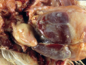
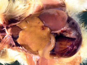
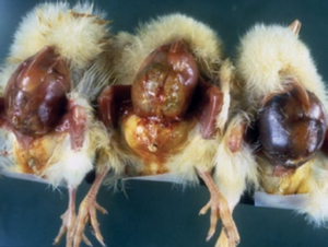
Yolk Sac infection (Mushy Chick Diseases, Omphalitis)
Subsequentlyafter hatching, during the first week this illness is one of the most common reasons for mortality in chicks. Clinical signs shown by E.coli infected chicks are like distended abdomen with the thickened prominent and necrotic navel. The chicks were dull and depressed huddle together. Brooding surrounding chick showed considerablemortality. Grossly, on opening of the dead birds theliver and kidney seen tinted, congested lungs, persistence of yolk whichcoagulated, inspissated andinvolvedwith intestine near the Meckel’s diverticulum. Due to fecal contamination of hatchery eggs, E.coli infect these embryonated eggs and lodge in the intestine of newly hatched chicks. The bacteria multiply rapidly in intestine and it leads to severe spread of infections among chicks in hatchery and brooder whose environment has not sufficient humidity.
Coli Granuloma (Hjarre’s disease)
Adult hens which given contaminated water typically affected by this condition. Dull, depression, loss of condition this type of clinical signs showed by birds leads to there death. At necropsy granulomas are seen in intestinal serosa and in mesentery specially in caeca. Often the affected liver is hard, mottled, discolored and puffy. histopathologically there is infiltration of inflammatory and occasionally giant cells in the intestinal necrotic mass. There is no effective method for control or treatment. The deprived environmental settings in small flocks activate recurrentE.coli infection which can be checked by proper training and expert guidance.
Swollen head syndrome
In broiler breeders and commercial layers, oedematous swelling over the eye is characteristic sign of swollen head syndrome. The lesions comprising of, facial oedema, caseous exudate in the conjunctiva, and lachrymal gland. Other viruses and primary infection predisposing these diseases leading to the syndrome. Antibacterial medication has beenreported to control the disease.
 |
 |
 |
||||||
|
|
|
||||||
- Fowl Typhoid
Fowl typhoid is a septicemic disease of domesticated bird causing heavy mortality,depends upon virulence of the organism usually affecting growers or adult birds. Etiological agent of this disease is Salmonella gallinarum(1.0–2.0 x 1.5μm) which is a nonmotile, Gram-negative bacillus. The organismclassified within group D of the Kauffmann–White scheme, having somatic antigen 0, 1, 9, 12. It can fermentlactose, sucrose, mannitol, maltose and dulcitol but does notferment lactose, sucrose and salicin. It grows on MacConkey and Blood agar as opaque glistening colonies.
Pathogenesis
The infected bird spread the infection through droppings, as it persists in faeces for at least a month.Transmission ofS. gallinarum occurs both vertical and horizontal root.Horizontal infection occurs through ingestion of faecal contaminated food and water. In diseased carcasses the organism survives for much longer periods from where rats, dogs and wild birds spread this disease flock to flock. Recovered birds acts as a carrier for further spreading of diseases. Vertical spread through egg can infect breeder, layer and broiler farmers.Similarly, attendants, visitors, etc. may carry infection from one farm to another,or house to house, unless appropriate disinfection procedures are in place.
Clinical signs
Generally, outbreaks of fowl typhoid seen at the end of summer season, when the weather is wet and moist. There is increase in mortality tailed by a drop in food consumption and drop in egg production. Ruffled feathers, dullness with close eyeand depression are common feature in infected birds. The characteristic clinical signis a watery to mucoid yellow diarrhea, occasionally respiratory distress with gasping can occur.In chronic phase there is loss of body condition and due to intense anaemia which results in to shrunken, pale combs and wattles. Due to shorter incubation period (4 to 6 days)the disease will spread quicklyover the flock andcan result in 100% morbidity and 50% or more mortality. Vertical transmission through Egg leads to an increase in dead-inshell embryos and small, weak, moribund or dead chicks on the hatching trays. Signs are nonspecific in affected chicks showing Yellow, pasty droppings, weakness, reluctance to move, tendency to huddle and a drop in food consumption. Occasionally there is respiratory distress with fast breathing and wheezing.
Lesions
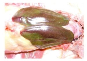
As this issepticemic disease, subcutaneous and skeletal vessels are prominent, congested and dark in colour. The Liver is invariably enlargedshows typically greenish bronze appearance which is an consistent finding. The spleen likewise found enlargedalso there is a catarrhal enteritis contain viscous, slimy, bile-stained material, mostly involving the small intestine, is usually present. Dark-brown bone marrow, an additional characteristic feature seen in this disease. Emaciated, anaemicwith focal necrosis in the heart, intestines, pancreas and liver arefound chronic phase of fowl typhoid. Pericarditis with deposition of the fibrin on the surface of the heart along with infiltration of yellow fluid in the pericardial sac is a feature of chronic fowl typhoid. Large greyish white nodules (Salmonella bumps) bulge out from the surface of the cardiac muscles. In young chicks, additionally there are necrotic foci in the lungs and gizzard. In layers, hemorrhagic, misshapen and rupture ova cause peritonitis. The ovaries are pedunculated with misformed follicle. In the testicles of the fowl typhoid affected cockerel’s well-defined necrotic foci have been reported.
Diagnosis
Isolation and identification of S. Gallinarumwhich grows on blood agar with opaque, glistening colonies and will growreadily on MacConkey agar as pale, non-lactose-fermenting colonies. The rapid plate agglutination test can be used. ELISA has also been demonstrated to be of use as a flock test.
Treatment
A number of antibacterial agents used like Furazolidone given continuously in the feed for 10 days at a level of 0.04% was generally considered to be the best treatment regimen. Reinfection of susceptible birds with the disease recycling in the flock also occurs. Therefore,its always preferred culling of chronically affected birds and reactors to a blood test and the prompt removal and incineration of all carcasses. Such resistance is not readily detected by disc diffusion tests.
Vaccination
Live attenuated rough strain ofS. Gallinarum known as vaccine 9R given subcutaneously inchickens between 10and 18 weeks of age it usually gave solid, long-lasting immunity. Vaccinated birds can carry and excrete the rough strain ofS. Gallinarum for long periods, and it can also be transmitted through the eggs laid by vaccinated birds.
|
||||||||||||
|
||||||||||||
|
||||||||||||

- Paratyphoid Infections (Avian Salmonellosis)
Salmonella received international attention because of their role in foodborne outbreaks of human illness by means of its involvement in food vehicles contaminating eggs and meatof poultry birds. At present more than 2500 different serovars have been identified, described and reported in poultry. Salmonella species has wide host range and the pathogenic syndrome is different for different species.Out of these Salmonella species 10 to 12 serovar is responsible for poultry diseases. Fowl typhoid infections is most important bacterial disease of hatcheries result in to huge mortality. Because of its chronic nature and difficulties in eradication Salmonellosis has huge impact on the economy of poultry industries of India. Paratyphoid infection has stuntingand debilitating effect on bird making them prone to many other infections.Salmonella genus are Gram-negative, non-capsulated, nonsporing (2–4 x 0.5μm)flagellated rods. Salmonellas are facultatively anaerobic, can cultivate on beef infusion agar forming thick, greyish-white, dome-shaped colonies. They all can survive for several months away from the host and can ferment glucose but notlactose, all reduce nitrates to nitrites.
Pathogenesis
After ingestion organism may colonize theintestine leading to formation of clinical signs, but if bird recover it recovers within 60 days. However, infected birds become carrier after recovery andshade the bacteriumfor a long time.Salmonella typhimurium and Salmonella enteritidiswhich are enterica serovars can easily introduced into poultry farms through rodents, insects, birds or fomites as it is profusely present in the environment. Newly hatched chicks get Infection as a result of massive multiplication within the alimentary tract which results in extensive infection within the house or hatchery where birds are transferred.Invasion to the spleen hampers the immunity and may results in systemicspread and multiplication. Certain strains like S. Enteritidisinfect reproductivetract, especially oviductled to vertical transmission. Salmonella serotypes produce endotoxins which are responsible for production of thelesions and mortality generally in first two weeks after hatching. Feed and water are contaminated by feces of brooder bird and because of that, infection rapidly transmitted to young birds by inhalation or by direct consumption. Frequent passage of the bacteriaconsequencestosurge of infection severely.
Signs
Hatching eggs infected with Salmonellosis sometimes shows dead embryos. Clinical signs like ruffled feathers, droop down head and closed eyes. There is pesty diarrhoea which soiling the vent, anorexia and reduce in water intake. Commonly near to the brooder bird huddle together. There is blindness because of corneal opacity.
Lesions
Cracas are emaciated, dehydrated, yolks are coagulated, congestion in liver and spleen with necrotic foci, pericarditis and congested kidney, grossly this type of lesions are seen at postmortem in birds died because of salmonella.
Diagnosis
Diagnosis can be done using clinical signs and symptoms like reduced body weight in broilers and growers and drop in egg production. Isolation and identification of organism from intestine specially from caeca, cloacal swabs, dead embryo eggs and litter. By using ELISA antibodies of variety of Salmonella antigen can be monitored.
Control
Antibacterial such as amoxicillin, tetracyclines, potentiated sulphonamides, spectinomycin, enrofloxacin and other fluoroquinolones are useful to limit morbidity and mortality within a flock. Along with this many effective biosecurity programme have to done to prevent infection in the first place. Proper staff hygiene on a poultry farm should be maintain, who may carry Salmonella organisms mechanically by means of contaminated footwear, clothing and hands.
Litter contamination specially by young chicks should be minimize as this is the prime source of infection to spread. Rodents, wild birds, insects etc, who can transfer or contaminate the flock in farm, feed, water should be checked. Effective disinfection of poultry houses, surrounding environment and equipment is crucial to prevent any carry over of salmonellas to the new flock. WHO has provided guidelines on cleaning, disinfection and vector control in Salmonella-infected
poultry flocks, with special reference to S. Enteritidis.Active monitoring of flock is essential at all stages of production to detect infections and to establish proper control procedures. Constant vigilance is desired to continue the maximum standards of management and disease security at breeding farms, hatcheries, rearing farms, feed mills and processing plants in order to prevent the introduction and spread of salmonellosis, both from established serovars but also from new oremerging strains.
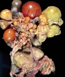
Figure 9 Enlarged and congested spleen and liver with pale yellow fibrinonecrotic cast in the lumen of cecum in a 10-day-old chick |
|||||||||

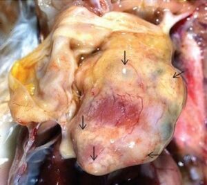
Figure 8 Ovary with multiple misshapen gray nodules |
|||||||||
|
|||||||||
Final Conclusion:
Bacterial diseases have devastating effects on the Poultry Industryand on poultry production systems. They cause mortality, reduced growth rates, decreased egg production, and condemnation of meat and eggs due to contamination. Additionally, outbreaks of zoonotic bacterial diseases, such as salmonellosis, can tarnish the reputation of the poultry industry and pose risks to public health.By implementing comprehensive prevention and control strategies, poultry producers can mitigate the risks posed by bacterial pathogens and ensure the sustainable production of safe and wholesome poultry products. Collaboration between industry stakeholders, veterinary professionals, and regulatory agencies is essential to address the complex challenges associated with bacterial diseases in poultry and safeguard the integrity of the poultry supply chain.