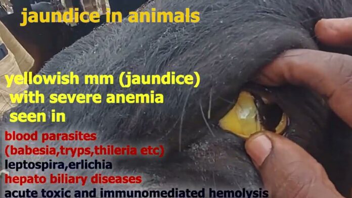
JAUNDICE (Icterus) IN ANIMALS
Dr. Aruna Maramulla
Contract teaching faculty, Department of VCC, College of veterinary science, Mamnoor, P.V Narasimha Rao Telangana Veterinary University, Hyderabad, Telangana.
Definition: Jaundice means yellowish discolouration of all visible mucus membranes, body tissues and body fluids i.e., both secretions and excretions.
Classification:
It is classified into three forms:
1) Prehepatic /Haemolytic jaundice
2) Hepatic /Toxic jaundice
3) Posthepatic/obstructive jaundice.
Prehepatic / Haemolytic jaundice:
It is due to excessive lysis of RBCs and occurs prior to the passing of blood to the liver so-called prehepatic jaundice
- Haemoprotozoan infections: Theileriasis, Babesiosis, Anaplasmosis
- Bacterial infections: Leptospirosis, Bacillary haemoglobinuria
- Viral infections: Equine infectious anaemia
- Deficiency diseases: phosphorus deficiency, Cu deficiency
- Metabolic diseases: postparturient haemoglobinuria
- Poisonings: snake bite, copper poisoning, onion poisoning, water intoxication
- Isoimmune haemolytic anaemia
Pathogenesis:
Excessive lysis of RBC
¦
Excess formation of Haemobilirubin (indirect unconjugated)
¦
The inability of the liver to convert excessively formed haemo bilirubin to chole-bilirubin
¦
Rise in haemobilirubin (indirect) level in blood leads to increased levels of cholebilirubin (direct)
¦
Accumulation of Excess bilirubin in the Intestine
¦
More formation of Stercobilinogen and Urobilinogen and the appearance of icterus visible mucous membranes
- II) Hepatic /Toxic Jaundice: It occurs due to damage to hepatic cells and usually due to certain toxins and hence also called as toxic jaundice.
Causes:
Toxins: (hepatitis)
Inorganic – (Cu, P, As, Pb, Hg, CCl4 etc.), Organic – (Gossypol, coal tar, alcohol etc), Plants – (Senecio, Crotalaria, Fribulus, Lantana), Fungi – Aspergillus, penicillum, fusarium, Algal toxins, Drugs- (Tetracycline, paracetamol etc).
Infectious agents
Bacteria – (Leptospira, Clostridium novyi, E-coli, Salmonella, Septicemic listeriosis),Virus – (Equine infectious anaemia, Infectious canine hepatitis, Feline panleukopenia), Chlamydia – Rift valley fever, Parasitic causes: Acute /chronic liver fluke infestation, migrating larvae of Ascaris spp, Nutritional causes – (Deficiency of methionine and choline, vit E and selenium), Congestive heart failure leading to increase in hydrostatic pressure in the sinusoids of the liver, Miscellaneous causes – Indirect hepatic damage caused by Diabetes mellitus, ketosis, pregnancy toxaemia, fatty cow syndrome.
Pathogenesis:
Haemolysis is normal &Haemobilirubin formation is normal. Due to liver damage liver cells are unable to convert normal amount of haemobilirubin to cholebilirubin. Cholebilirubin enters in blood circulation Via. hepatic circulation. Increased level of bilirubin in blood, urine causes ictric mucous membranes and dark yellowish urine. Decreased level of cholebilirubin in GIT leads to low bilirubin levels in GIT which exbit in form of light yellowish feces.
3) Obstructive/ post –hepatic jaundice: It is due to obstruction of bile duct and defect in bile duct which is next in sequence to liver so it is also called post – hepatic jaundice
Causes: It is due to obstruction of bile duct caused by Cholangitis (Inflammation of bile duct), Cholecystitis (Inflammation of gall bladder), Cholelithiasis (gall / bile stones), Parasites e.g Dicrocoeilium, Toxocara, Mature liver flukes etc. Neoplasms cysts, Abscess in bile duct or exterior to bile duct which may cause complete or partial obstruction.
Pathogenesis:
RBC lysis, Haemobilirubin, Cholebilirubin formation are normal but obstruction of bile duct results in back flow of cholebilirubin which enters in blood circulation.
Increase in level of cholebilirubin in blood leads to dark yellow straining of mucus membranes
Absence/decrease level of cholebilirubin in GIT causes decrease formation of stercobilinogen which appear as white/ chalky/ clay colour feces.
Increase excretion of cholebilirubin in urine causes intense yellow colour urine
Clinical signs:
Yellowish discolouration of mucus membrane (markedly visible in sclera). Light yellow, dark yellow or greenish yellow urine. Red urine in haemolytic jaundice. Dark yellow to clay colour faeces (hypopigmentation) faeces may contain fat i.e steatorrhoea. Muscular weakness. Mental depression. Loss of body weight (emaciation). Loss of appetite. Vomition in dogs and pigs. Constipation followed by diarrohea. Severe anaemia in haemolytic jaundice. Sometimes oedema, bottle jaw, ascites. Excitement, coma and death.
Diagnosis:
Differential Diagnosis of jaundice:
| S.No | Parameter | Haemolytic | Toxic | Obstructive |
| 01 | Erythrolysis | Increased | Normal | Normal |
| 02 | Liver | Normal | Normal | Normal |
| 03 | Bile duct | Normal | Normal | Abnormal |
| 04 | Serum indirect bilirubin | Very high | Increased | Normal |
| 05 | Serum direct bilirubin | Normal | Increased | Very high |
| 06 | Urine bilirubin | Absent | Increased | Very high |
| 07 | Urobilinogen | Increased | Normal to decreased | Decreased to absent |
| 08 | Urine colour | Light yellow | Intense | Intense |
| 09 | Stercobilinogen | Increased | Normal to decreased | Decreased/absent |
| 10 | Colour of faeces | Hyperpigmented | Normal to pale | Hypopigmented chalky / clay colour |
| 11 | Colour of mucus membrane | Slight to moderate yellow | Moderate yellow | Intense yellow |
| 12 | Colour of serum | Slight to moderate yellow / reddish | Slight to moderate yellow | Intense yellow |
| 13 | Icterus index (8.4 – 9.8 normal in cattle) | Low or moderate | Moderate | High |
| 14 | Vanden Bergh’s test | Indirect | Biphasic | Direct |
| 15 | Liver function test. | Negative | Positive | Negative (initially) |
- II) Diagnosis
Liver function tests: Vanden Berghs reaction – Indirect, direct or biphasic. Increased blood clotting time and Serum bilirubin levels.
Urine analysis: Urine samples are positive for bile pigments and bile salts.
General care: Give complete rest, provide fat and salt free, carbohydrate rich, high quality protein diet.
Specific treatment:
- Antibiotics orally of parenterally e.g Amoxycillin/Ampicillin @ 5-10mg /kg IM, IV, PO.
- Antiprotozoan drugs: e.g. Berenil @ 5.5-7.0mg /kg IM
- Antihelmintics: g. Flukicides distodin 10-15 mg /kg PO.Fenbendazole 50-10mg /kg PO
- Surgical intervention in gall stone, tumor, neoplasm etc.
Supportive treatment:
- Dextrose 5% or 10% orally/parentrally
- Liver tonics like Belamyl @ 5-10ml IM in larger animals, Neohepatex @ 1-2ml IM in dogs, Livogen /liv 52 syrup orally.
- Steroids: e.g. Dexamethasone @ 0.04 mg /kg IM
- Lipotrophic agents e.g. choline, methionine
- Calcium therapy to prevent guanidin intoxication @ 10ml IV as 10% solution in dog
- Mild purgatives in toxic jaundice e.g., MgSO4 @ 250-350 gm orally in large animals.
- Diuretics in ascites /edema e.g .ridema /lasix ( furasemide ) @ 1-2 mg/kg IM /IV.


