Johne’s disease (Paratuberculosis): Upcoming Zoonoses
The zoonotic significance of paratuberculosis has been debated for a long time (Dalziel 1913). After the identification and isolation of Mycobacterium avium paratuberculosis (MAP) in humans suffering from Crohn’s disease in 1986 (Chiodini et al. 1986), the attention of many researchers diverted to finding the zoonotic potential of paratuberculosis (Grant 2005). Furthermore, MAP is considered as an important agent in the pathogenesis of certain human auto-immune diseases. These diseases include human immunodeficiency disease, diabetes mellitus, encephalomyelitis disseminate, chronic autoimmune thyroiditis, Besnier-Boeck-Schaumann disease (Sechi et al. 2008; Paccagnini et al. 2009; Sisto et al. 2010; Cossu et al. 2013). The zoonotic potential of paratuberculosis is based on the analogical relationship of clinical symptoms between Johne’s disease in animals (i.e. both ruminants and nonruminants) and Crohn’s disease in human beings (El-Zaatari et al. 2001).
Paratuberculosis, also known as Johne’s disease, is a chronic, contagious bacterial disease of the intestinal tract that primarily affects sheep and cattle, goats as well as other ruminant species. The disease has also been reported in horses, pigs, deer, alpaca, llama, rabbits, stoat, fox, and weasel. The disease is caused by a bacterium called Mycobacterium avium subsp. paratuberculosis (MAP). Disease has long and protracted incubation period which may extend even up to 2 years or more.
Economic importance
Johne‘s disease has worldwide distribution and it has been increasing range of animal species. In India, JD has been endemic and highly prevalent. It has devastating effects on livestock sector in terms of production and economics, where losses occur due to subclinical stage of disease, in the form of premature culling, reduced carcass value, reduced weight gain, increased susceptibility to other infections, reduced fertility, reduced feed efficiency, reduced milk yield, reduced salvage value at slaughter, increased treatment costs and shorter life expectancy.
Transmission
In ruminants, MAP is mainly transmitted by the fecal-oral route. Infected animals can shed large numbers of organisms in the feces. Young animals are most susceptible to infection and usually become infected when they nurse from an udder soiled with feces or housed in contaminated pens. They may also be infected when they drink milk or colostrums from infected dam. Little is known about the transmission of MAP in nonruminant species, but fecal-oral spread is likely to be important. Predation might be a route of transmission to carnivores or omnivores.
Clinical signs
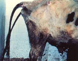
Clinical signs usually first appear in adulthood, but the disease can occur in animals at any age over 1-2 years and in dairy cattle is most frequently reported in the 3-5 year old age group. Clinically infected animals show watery diarrhea, emaciation and eventually death due to lack of effective treatment. The initial symptoms can be subtle and may be limited to weight loss, decreased milk production or roughening of the hair coat. The diarrhea is usually thick, without blood, mucus or epithelial debris, and may be intermittent at first. As the disease progresses, the diarrhea becomes more constant and severe over weeks or months and intermandibular edema may occur. The temperature and appetite are usually normal and animals are alert. The clinical signs are similar in other ruminants. In sheep and goats, the wool is often damaged and easily shed, and diarrhea is less common than in cattle.
Lesions
In cattle, early lesions occur in the walls of the small intestine and the draining mesenteric lymph nodes and infection is confined to these sites at this stage. As the disease progresses, gross lesions occur in the ileum, jejunum, terminal small intestine, caecum and colon. The walls of intestine become 2-20 times thickened. The mucosa of intestine is folded showing transverse corrugations (Chacon et al., 2004). Similar lesions occur in sheep and goats. The mucosa is often only slightly thickened in these species, but caseated or calcified nodules are sometimes found in the intestine and associated lymph nodes. The lesions in other ruminants resemble those found in cattle, sheep and goats.
Zoonotic Risk
MAP is an emerging pathogen of global concern. However, limited data suggest that MAP may be involved in Crohn’s disease (CD), chronic granulomatous enteritis of humans that resembles Johne’s disease (JD) and share certain clinical and histopathological similarities with JD in animals (Pickup et al., 2005). CD is characterized by periods of malaise, abdominal pain, chronic weight loss and diarrhea, with remissions and relapses. The disease often begins between the ages of 16 and 25 years, and persists lifelong. There is no cure. The cause of Crohn’s disease is unknown; however, it may be the result of several interacting factors including a genetic predisposition, an abnormal immune response, and environmental factors including responses to intestinal microorganisms. MAP has been found in some CD patients; however, isolation is rare and studies to date have not been able to determine whether this organism has a causative role or is simply an “innocent bystander” that can grow in the inflamed intestinal wall. However, MAP is consistently detected by PCR in people with Crohn’s disease. This fact, coupled with its broad host range, including nonhuman primates, indicates that paratuberculosis should be considered a zoonotic risk until the situation is clarified (Mishina et al., 1996).
Diagnosis
Diagnosis of MAP is challenging due to complex pathogenesis, long incubation period, and intracellular location of pathogen. Early identification of infected animal is essential to prevent further spread of the disease (http://www.nap.edu/books/0309086116/html). Early “silent” infections can be detected only by culturing the organisms from postmortem tissues or, rarely, by histopathology. Subclinical carriers can be identified with serology, delayed-type hypersensitivity (DTH) reactions, polymerase chain reaction (PCR) assays or fecal culture (Amarapurkar et al., 2004).
Microscopy
Ziehl-Neelsen stains can be used to detect MAP in the feces, smears from intestinal mucosa or the cut surfaces of lymph nodes; clumps of small, strongly acid-fast bacilli are diagnostic.
Culture
Bacteria can be cultured from the feces, thickened areas of the intestinal wall, and ileal, mesenteric and ileocecal lymph nodes. Suitable media include Herrold’s egg yolk medium, (HEYM) modified Dubos’s medium and Middlebrook 7H9, 7H10 and 7H11 media.
Serological diagnosis
Serology can be used for the presumptive identification of infected animals, as well as to estimate the prevalence of infection in a herd or confirm paratuberculosis in animals with clinical signs. A variety of serological tests are available, including complement fixation, enzyme-linked immunosorbent assays (ELISAs) and agar gel immunodiffusion (AGID).
Tests for cell mediated immunity
Intradermal testing with johnin or avian purified protein derivative has been used widely to detect delayed-type hypersensitivity (DTH) reactions to MAP; however, this test is insensitive and nonspecific reactions are common. DTH reactions may diminish or disappear as the disease progresses. The test is carried out by the intradermal inoculation of 0.1 ml of antigen into a clipped or shaven site, usually on the side of the middle third of the neck. The skin thickness is measured with calipers before and 72 hours after inoculation. Increases in skin thickness of over 2 mm should be regarded as indicating the presence of DTH. In vitro tests that detect cell-mediated immunity to MAP include a gamma interferon assay (IFN-γ) and a lymphocyte transformation test (LTT). The IFN-γ assay is a sensitive laboratory test, but less useful and costly. The cell-mediated immune (CMI) response elicited in the early stage of infection is detected by the release of IFN-γ in the blood from sensitized lymphocytes during 18-36-hour incubation period with specific antigen.
PCR and DNA probes
PCR and DNA probes are used to detect MAP and distinguish it from other species and subspecies of mycobacteria.
Prevention and control
Prevention & control of Johne’s disease is important as treatment is not available. Control involves good sanitation and management practices including screening tests for new animals to identify and eliminate infected animals and ongoing surveillance of adult animals. In herds affected with paratuberculosis, calves, kids, or lambs should be birthed in areas free of manure, removed from the dam immediately after birth, bottlefed pasteurized colostrum and raised separate from adults until at least one year old. This reduces the chance of transmission of disease to this most susceptible population. Also, reducing fecal contamination in animal housing areas by elevating food and water sources is recommended. There are some vaccines for this disease; however they are used only in very well defined situations and under strict regulatory control. Vaccination of young calves has shown a reduction in disease incidence but it does not prevent shedding or subsequent new cases in the herd. However, vaccination may interfere with eradication programmes that are based on detection and subsequent elimination of infected animals. Vaccination against Paratuberculosis can also interfere with tests for bovine tuberculosis. Strategies to control this disease include improved management practices, testing and culling and vaccination.
CONCLUSION
The diagnosis of paratuberculosis can provisionally be made on clinical grounds but confirmation requires certain laboratory investigations. Isolation of the organism is the gold standard diagnostic test. Several serological techniques are employed in the diagnosis of paratuberculosis. Molecular tests have recently been utilized in the diagnosis of the disease. The knowledge of how MAP causes disease still lags behind than for other pathogenic bacteria. Johne‘s disease control programs are the immediate requirements of the country in order to boost per animal productivity. The potential role of MAP in the etiology of Crohn’s disease deserves substantial future investigation.
Compiled & Shared by- This paper is a compilation of groupwork provided by the Team, LITD (Livestock Institute of Training & Development)
Image-Courtesy-Google
Reference-On Request


