


Lumpy Skin Disease- Its Epidemio-Pathology and Management
Das T1, Das NK2 and Rout, BR3
1Scientist, ICAR-DFMD, Bhubaneswar
2BVO, Bisoi, Mayurbhanj, FARD, Government of Odisha
3BVO, Khandapada, Nayagarh, FARD, Government of Odisha
Corresponding author E mail id: tarenisahoo@gmail.com
Our country, India is having world’s largest livestock population with 192.49 million cattle and 109.85 million buffalo population (20th Livestock Census). It plays an important role in Indian economy as 20.5 million people depend on this sector for their livelihood. The most important obstacle to the growth of livestock industry is morbidity and mortality associated with various emerging and re-emerging diseases. Recently, Lumpy skin disease has been reported to be a major health issue and associated with huge economic losses in many developing countries including India. It is a vector borne emerging viral transboundary disease of high economic importance affecting cattle and water buffaloes. The virus causes both acute and subacute disease in the susceptible hosts of all ages and breeds. But young and first lactating cattle are more frequently affected. It is having a substantial economic impact on large animal industry due to decreased milk and meat production, decreased fertility in both male and female animals, abortions, damage to hide and skin, restriction on trade and movement of animals, high cost in disease treatment and prevention, mortality of affected animals etc. The present article describes the epidemio-pathology and management of lumpy skin disease.
Etiology
Lumpy skin disease (LSD) virus belongs to Genus Capri poxvirus under family Poxviridae (Gupta et al., 2020). The prototype strain of the virus is known as Neethling Pox virus. It is an enveloped ds DNA virus with 151kbp genome and of relatively large size (320nm X 260nm) and multiplies in the cytoplasm of the host cells. It encodes 30 proteins which are antigenically closely related to sheep pox and goat pox virus. The virus is stable for a long period in the ambient temperature and can persist for many months in the dark environment like animal shed. It can persist for 18 days, 33 days 35 days and up to 6 months in the dried hides, necrotic nodules, dried skin crusts and fomites respectively. The virus is susceptible to high alkaline and acid PH and can be inactivated at 550 C for 2 hours and at 650 C for half an hour. The virus can be inactivated by chemical compounds like 20% ether, chloroform, 0.5% quaternary ammonium compound, 1% formalin, 2-3% Na hypochlorite, iodide compounds etc. (Gupta et al., 2020).
Epidemiology
The disease was first reported in Zambia in 1929 which was thought to be pseudo urticaria caused by biting insect (McDonald, 1931). The disease then spread rapidly and extensively across Africa. It was then reported in Egypt, the Middle East, South East Europe, West Asian region, Central Asia, the Balkans, the Caucasus and more recently in South Asia, China, Bangladesh, Nepal, Thailand etc. (OIE, 2020; Anwar et al., 2022). During the period from 2005 to 2020, Africa had highest outbreak reports (29, 966), then followed by Asia (8837) and Europe (2471). In Africa, Zimbabwe and Ethiopia; in Asia, Oman, Turkey, Iran and Thailand; in Europe, Russia had reported highest numbers of outbreaks (Anwar et al., 2022). In India, the disease was first reported in five districts of Odisha in August, 2019 with 7.1% morbidity and no mortality (Sudhakar et al., 2020). LSD has been reported in many states of India including Maharashtra, Madhya Pradesh, Gujrat, Goa, Karnataka, Kerala, Andhra Pradesh, Odisha, West Bengal, North East States, Jharkhand, Chhattisgarh and more recently in Punjab.
The disease is host specific affecting cattle and water buffaloes. The disease has been demonstrated in wild animals like Arabian Oryx, Springbok, giraffe and impala experimentally, not naturally. No evidence of the disease has been reported in small ruminants like sheep and goats naturally and the virus does not affect humans because of its non-zoonotic nature (Tuppurainen et al., 2017). Breed wise, cross breed animals were found to be more affected than indigenous zebu cattle. The fine skinned breeds like Holstein Friesian and Jersey breeds are more susceptible. Age wise, the younger animals and the animals on their first lactation are more severely affected than others (Kayesh et al., 2020; Mahor et al., 2022).
The virus is excreted through skin scab, blood, milk, semen, nasal, lachrymal secretion and saliva. The LSDV is mainly transmitted by arthropod vectors with more incidence in wet season because of abundant biting fly population. Both transstadial and mechanical transmission have been reported. The various vectors like Stomoxys calcitrans, Culicoides nubecolosus and Aedes aegypti are potentially efficient transmitter of the virus (Anz-Bernardo et al., 2021). Ticks like Rhipicephalus species and Amblyomma species have shown to transmit LSDV. Rarely, the virus is transmitted via direct contact and contaminated feed and water. Recently, the evidence of naturally occurring indirect contact mode transmission of vaccine derived virulent LSDV strain was reported (Aleksandr et al., 2020).
The morbidity and mortality rated in LSD depend on several factors like nutritional, health, immune status and breed of cattle, population and dissemination of arthropod vectors, virulence of virus, climate, geography and managemental factors. The morbidity rate varies from 5-45% and the mortality rate is less than 10% (Chaoudhari et al., 2020).
Pathology
After subcutaneous or intradermal inoculation of the LSDV virus, a localized swelling at the site of entry develops. The virus multiplies and causes enlargement of regional lymph nodes and lymphadenitis. Then viraemia and febrile reactions occur which persists for 15 days. The virus replicates inside the host cells such as macrophages, fibroblasts, pericytes and endothelial cells leading to vasculitis and lymphangitis, consequently thrombosis and infarction develop resulting in oedema and necrosis. The virus multiplies in the keratinocytes resulting in hyperplasia and ballooning degeneration, vesicles and ulcers formation and infiltration of inflammatory cells in the dermis. Development of inflammatory nodules in the skin takes 7-19 days after viral inoculation. After recovery from clinical disease, a lifelong cell mediated immunity (CMI) develops and calves borne to these infected animals obtain maternal antibodies which can protect them up to 6 months period (Ahmed et al., 2020).
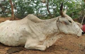
The incubation period varies from 1-2 weeks in experimental infection and 2-5 weeks in natural infection. The clinical signs include initial pyrexia which may exceed 410C and persists for 1 week. Then inappetence, salivation, nasal and ocular discharge, enlarged superficial lymph nodes, drastic reduction in milk yield, loss of body condition etc. are observed. Cutaneous lesions appear typically on the neck, head, leg, tail, back, udder, scrotum, vulva, perineum etc. The lesions are classically multiple, round, irregular, firm, circumscribed, coalescing, slightly raised nodules of 2-7 centimeter in diameter (Fig. 1). The lesions involve entire thickness of skin including epidermis, dermis, subcutis and rarely involves adjacent muscles. On the cut sections, nodules are cream grey to white in color with oozing of serum initially. Afterward, the nodules harden and form necrotic plug/dimple/sit fast in the center. It may peel off leaving a hole in the center which may be contaminated with secondary bacteria or infested with fly worms. The edematous swelling develops on face, brisket and limbs leading to lameness. The necrotic and ulcerative lesions also develop on the muzzle, lips, gingiva, dental pad, larynx, trachea, fore stomach, abomasum etc. which leads to development of gastroenteritis, stenosis of trachea and pneumonia. The affected animals may suffer from anestrous, infertility, orchitis, mastitis and abortion. The severely affected animals may die and the recovered animals suffer from weakness and debility for 6 months (OIE, 2017; Ahmed et al., 2020). The histopathological lesions consist of proliferation of epidermis with rete peg formation associated with vacuolation of epidermal cells with presence of numerous eosinophilic intracytoplasmic inclusion bodies. The inclusion bodies may be also observed in the endothelial cells, fibroblasts, macrophages, pericytes and keratinocytes. In many cases, In the dermis, vasculitis with fibrinoid necrosis, lymphangitis, thrombosis, infarction, oedema, infiltration of inflammatory cells including large epithelioid macrophage like cells, separation of dermis and epidermis are observed (OIE, 2017).
 |
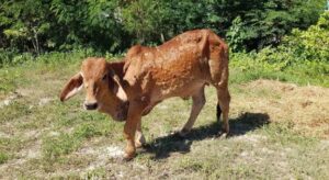 |
| Fig. 1. Multiple, round, irregular, firm, circumscribed, slightly raised nodules distributed all over the body in cattle affected with LSD | |
Diagnosis
The LSD can be diagnosed based on characteristics clinical signs and lesions like high fever, ocular and nasal discharge, multiple typical skin nodules (10-50 mm diameter), markedly enlarged prescapular and pre femoral lymph nodes etc. and based on incidence of the diseases.
In early stages, skin biopsies can be preserved in 10% buffered formalin and can be routinely processed for histopathology and stained with hematoxylin and eosin stain. The typical skin lesions like ballooning degeneration, presence of typical intracytoplasmic inclusion bodies etc. are helpful in diagnosis.
For virus isolation, tissue samples like skin nodules (preferably should be collected within 1 week of observation of clinical signs) and blood samples collected in heparin or EDTA during viremic stage are generally used. It is a golden standard but time-consuming method for LSDV diagnosis. Primary or secondary culture of bovine dermal cells, lamb testis, African Green Monkey Kidney, Madin- Darby Bovine Kidney cell line (MDBK), chorio allantoic membrane (CAM) of embryonated chicken eggs etc. are used for isolation LSDV. The characteristics CPE in culture include retraction of cell membrane, cell rounding, chromatin margination and development of intracytoplasmic inclusion bodies. In CAMs, characteristics pin head to pin point sized pock lesions are reported after LSDV infection (OIE, 2017; Amin et al., 2021). Transmission Electron microscopy can be used to visualize typical virions. Serological assays like serum neutralization tests (SNT), immune peroxidase monolayer assay (IPMA), agar gel immunodiffusion test (AGID), western blot analysis and indirect fluorescent antibody test (IFA) are suitable methods to investigate recent outbreaks and disease-free status of the country (OIE, 2017). Immunohistochemistry (IHC) using specific antibodies to detect LSDV antigens in tissue samples has been described (Amin et al., 2021). Molecular methods like PCR are used routinely for detection of LSDV DNA in blood, semen and tissue samples. It is a simple, fast and sensitive method. Another molecular method like loop mediated isothermal amplification (LAMP) has been described which is a simple, specific and cost-effective method for detection of LSDV (Mwanandata et al., 2018). A TaqMan probe based multiplex real time PCR has been validated for detection of wild type LSDV (Aggianiotaki et al., 2021). Currently, DIVA PCR kits are available for differentiating classical field strain and recombinant vaccine strain of LSDV (Byadovskaya et al., 2021).
Management (Treatment, control and prevention)
There is no specific antiviral drug for treatment of lumpy skin disease. The treatment is symptomatic and practiced to prevent secondary bacterial infection. The supportive care includes local dressing of skin with antiseptic cream to prevent fly infestation and secondary bacterial contamination, antibiotic treatment to prevent further bacterial infection and pneumonia, antihistamines and anti-inflammatory pain killers, antipyretic drugs, multivitamins, liver supportive therapy, intravenous fluid therapy etc. (Ahmed et al., 2020).
To prevent rapid and uncontrollable spread of LSD, effective control and preventive measures need to be implemented and followed. Early detection and rapid laboratory diagnosis are crucial for fruitful control of LSD. LSD awareness campaign should be carried out targeting farmers, veterinarians, Para veterinarians, drivers of cattle transport vehicles etc. for identifying infected animals and for informing to higher authorities. Also, recurrent sero-surveillance needs to be carried out in unnoticed outbreak to identify seropositive animals. Efficient vector control methods like limiting vector breeding sites, use of vector traps, use of insect proof housing for cattle and insecticide spray will reduce arthropod population, thereby reducing mechanical transmission of disease. The movement of infected animals should be restricted, the infected animals should be quarantined and the carcass should be properly discarded in order to prevent rapid and uncontrollable spread of the disease. Restriction to international trade of live animals and animal products should be imposed to prevent transboundary spread of the disease. The biosecurity should be enhanced in the infected farm and the farm premises should be properly disinfected with proper disinfectant and detergents. Newly purchased animals should be quarantined before introducing to healthy herds (Tuppurainen and Galon, 2016).
The most important and effective way to control LSD is effective and safe vaccination of regional cattle and water buffaloes against LSD. Several vaccines are available which provide protection against LSD to various level. Homologues live attenuated vaccine containing South African Neethling strain or KSGP O-240 or O-180 provides good protection to cattle against virulent field strain. As the members of Capri pox virus provide cross protection against each other, live attenuated sheep pox and goat pox vaccines are also used in cattle which provided good protection and seroconversion in cattle. Different strains like Gorgan goat pox strain, G2-LKW strain, Yugoslavian RM-65 Sheep pox strain, Romanian SPPV strain, Niskhi strain etc. are used in commercial vaccine production. Heterologous vaccines require low level of attenuation than homologous LSDV vaccine for safe use in cattle population. The potential problems associated with live attenuated vaccines are development of local inflammation or mild disease with skin lesions, risk of vaccine virus shedding and transmission, risk of development of recombinant strain, loss of disease-free status of corresponding vaccinated country etc. (Tuppurainen et al., 2021). Annual vaccination of animals older than 6 months should be carried out with use of single needle per single animal in order to prevent transmission of the disease (Mahor et al., 2022). Now a days, inactivated vaccines are also available in the market. Due to non-replicative nature of virus, it is safer to use than live attenuated vaccine. Inactivated Capri pox vaccine and inactivated oil adjuvanted vaccine based on Neethling Strain were developed, evaluated and produced good immune response (Hamdi et al., 2020; Wolff et al., 2020).
References
Agianniotaki EI, Chaintoutis SC, Haegeman A, De Clercq K, Chondrokouki E, Dovas CI. (2021). A TaqMan probe-based multiplex real-time PCR method for the specific detection of wild type lumpy skin disease virus with beta-actin as internal amplification control. Mol Cell Probes. 60:101778.
Ahmed N, Doley S, Barlaskar SA, Nath AJ, Yadav SN. (2020). Lumpy Skin Disease: an emerging bovine viral infection in India. Indian J. Anim. Hlth. 59(2):137-42.
Aleksandr K, Olga B, David WB, Pavel P, Yana P, Svetlana K, Alexander N, Vladimir R, Dmitriy L, Alexander S. (2020). Non-vector-borne transmission of lumpy skin disease virus. 10(1):7436.
Amin DM, Shehab G, Emran R, Hassanien RT, Alagmy GN, Hagag NM, Abd-El-Moniem MI, Habashi AR, Ibraheem EM, Shahein MA. (2021). Diagnosis of naturally occurring lumpy skin disease virus infection in cattle using virological, molecular, and immunohistopathological assays. Veterinary World. 14(8):2230.
Anwar A, Na-Lampang K, Preyavichyapugdee N, Punyapornwithaya V. (2022). Lumpy Skin Disease Outbreaks in Africa, Europe, and Asia (2005–2022): Multiple Change Point Analysis and Time Series Forecast. Viruses. 14(10):2203.
Anz-Bernardo B, Haga IR, Wijesiriwardana N, Basu S, Larner W, Diaz AV, Langlands Z, Denison E, Stoner J, White M, Sanders C, Hawes PC, Wilson AJ, Atkinson J, Batten C, Alphey L, Darpel KE, Gubbins S, Beard PM. (2021). Quantifying and Modeling the Acquisition and Retention of Lumpy Skin Disease Virus by Hematophagus Insects Reveals Clinically but Not Subclinically Affected Cattle Are Promoters of Viral Transmission and Key Targets for Control of Disease Outbreaks. J Virol. 95(9):e02239-20
Bhoopendra Singh Mahor, Amita Tiwari, Shivangi Udainiya, DK Gupta, Brejesh Singh and Ranbir Jatav. (2022). Lumpy skin disease: An economically devastating emerging viral disease. The Pharma Innovation Journal. 11(6S): 76-79.
Byadovskaya O, Pestova Y, Kononov A, Shumilova I, Kononova S, Nesterov A, Babiuk S, Sprygin A. (2021). Performance of the currently available DIVA real‐time PCR assays in classical and recombinant lumpy skin disease viruses. Transboundary and Emerging Diseases. 68(6):3020-4.
Choudhari AN, Moregaonkar SD, Gangane GR, Markandeya NM, Narladkar BW. (2020). Lumpy skin disease (lsd), an emerging disease in India: a review. Agricultural Reviews. 41(4):398-402.
Gupta T, Patial V, Bali D, Angaria S, Sharma M, Chahota R. (2020). A review: Lumpy skin disease and its emergence in India. Vet Res Commun. 44(3-4)
Hamdi J, Boumart Z, Daouam S, El Arkam A, Bamouh Z, Jazouli M, Tadlaoui KO, Fihri OF, Gavrilov B, El Harrak M. (2020). Development and evaluation of an inactivated lumpy skin disease vaccine for cattle. Veterinary microbiology. 245:108689.
Kayesh ME, Hussan MT, Hashem MA, Eliyas M, Anower AM. (2020). Lumpy skin disease virus infection: An emerging threat to cattle health in Bangladesh. Hosts and Viruses. 7(4):97.
MacDonald RAS. (1931). Pseudo – urticaria of cattle, North Rhodesia Department of Animal Health, Annual Report. pp20-21
Manual, OIE Terrestrial. (2017). Lumpy skin disease, Chapter 2.4. 13.
Mwanandota JJ, Macharia M, Ngeleja CM, Sallu RS, Yongolo MG, Mayenga C, Holton TA. (2018). Validation of a diagnostic tool for the diagnosis of lumpy skin disease. Vet Dermatol. 29(6):532-e178
Sudhakar SB, Mishra N, Kalaiyarasu S, Jhade SK, Hemadri D, Sood R, Bal GC, Nayak MK, Pradhan SK, Singh VP. (2020). Lumpy skin disease (LSD) outbreaks in cattle in Odisha state, India in August 2019: Epidemiological features and molecular studies. Transboundary and Emerging Diseases. 67(6):2408-22.
Tuppurainen E, Alexandrov T, Beltrán-Alcrudo DJ. (2017) Lumpy skin disease-a manual for veterinarians. FAO Animal Production and Health Manual.
Tuppurainen E, Dietze K, Wolff J, Bergmann H, Beltran-Alcrudo D, Fahrion A, Lamien CE, Busch F, Sauter-Louis C, Conraths FJ, De Clercq K. (2021). Vaccines and Vaccination against Lumpy Skin Disease. Vaccines. 9(10):1136.
Tuppurainen E, Galon N. (2016). Lumpy Skin Disease: Current situation in Europe and neighbouring regions and necessary control measures to halt the spread in South-East Europe. Europe–OIE Regional Commission.
Wolff J, Moritz T, Schlottau K, Hoffmann D, Beer M, Hoffmann B. (2020). Development of a safe and highly efficient inactivated vaccine candidate against lumpy skin disease virus. Vaccines. 9(1):4.