


Management of Coenurus gaigeri cyst in Sheep
Sudha Rani.R, Assistant Professor, SRDDL, IAH&VB, Hebbal, Bangalore-24
Abstract:
An two year male sheep was presented to an veterinary dispensary in Bangalore, Karnataka with the history of swelling in thigh region of the right hind leg. On physical examination, revealed a large fluctuating fluid filled sac with distinct dimension. The exploratory puncture of the swelling revealed a colorless transparent fluid confirming it as a cyst. On surgical removal of the cyst it appeared as transparent sac like structure with rice grain like particles attached to the sac. Parasitological examination of the sac revealed it as a Coenurus gaigeri, the intermediate stage of T. multiceps gaigeri. The animal recovered completely by 15th postoperative day without any complications.
Key words: Coenurus gaigeri cyst, sheep.
Introduction:
Coenurosis (gid or sturdy) is a fatal disease in sheep and goats, caused by larval forms of Taenia multiceps (Patro et al.1997). Whereas metacestodes of other cestodes of goat such as Coenurus cerebralis, Cysticercus tenuicollis, Cysticercus ovis, and hydatid cysts are more common. The cystic larvae (C. cerebralis) which develop in the brain or spinal cord will predominantly affect normal functioning of central nervous system (CNS) of the parasitized host. In recent studies various sites of prediction of the metacestode is reported in neck muscle, eyelid, skin, thigh muscles, abdominal muscles, heart, kidney and lymphnodes in goats (Varma et al.1994), with an alternate name (C. gaigeri). In reported cases of coenurie affected goats, the cysts anchor, develop, mature and cause asymptomatic focal lesions in various sites. The lesions persist throughout the life span of the host which is governed by multiple factors, including the quantity and periodicity of infection intake, subsequent host-parasite interaction, age and acquired immune status of the goat, etc. Chronic infections are more in aged between one to two years of goats. This article presents a case of C. gaigeri in the thigh muscle of a non-descript sheep and its successful surgical management.
Case history and Observations:
A 2yr old non descript male sheep was presented to an veterinary dispensary in Bangalore, Karnataka with history of swelling on posterior right hind leg at thigh region (Fig.1). The sheep had normal temperature, respiration and pulse. On physical examination revealed a large fluctuating fluid filled bladder with distinct dimension. On aspiration a clear fluid was observed and the fluid filled structure had a clear demarcation from its surroundings (Fig. 2). Based on clinical signs and physical examination, it was tentatively diagnosed as cyst. Surgery was done under local infiltration with 2 % lignocaine hydrochloride solution after aseptic preparation of the surgical site. Cyst was removed after separating it from muscular attachments (Fig. 3) and tincture of iodine was applied and the skin wound was closed by applying simple interrupted sutures. Post operatively the animal was given Inj. AC vet 100mg intramuscularly and liq. Fentas plus @ 1 ml/kg body weight orally. The sutures were removed on the next day and the cavities of the cysts were again cleaned and new sutures with betadine were applied (Fig.4). This procedure was followed on alternate days till the cavity of the cysts were obliterated (15 days). The Inj. AC vet was given for five days only to check secondary bacterial invasion. The animal was recovered fully by 15th post operative day.
Results and discussion:
On postoperative we observed the swelling subsided and the animal resumed normal posture and gait. The cyst was removed in intact and it was bladder like having containing colorless transparent fluid with numerous macroscopic invaginated protoscolices in clusters attached to the internal surface of its wall (Fig.3). On microscopic examination we visualized a single scolex with four suckers and one rostellum armed with a double crown of 30–32 hooks with hooklets. Typical taenid hooks were characteristics of coenurus and being it extra cranial, the cysts were identified as C. gaigeri, the intermediate stage of T. multiceps gaigeri.
Coenurosis (gid or sturdy) is principally a fatal disease of ungulates, especially sheep, caused by larval forms of Taenia multiceps (Varma et al.1989). The cystic larvae (C. cerebralis) which develops in the brain or spinal cord has predominantly affects normal functioning of the central nervous system (CNS) of the parasitized host including man (Sharma and Chauhan 2006). Apart from this, occasionally aberrant sites of prediction of the metacestode, especially in goats, with an alternate name (C. gaigeri), have been reported (El-Sinnary et al. 1999; Sharma and Chauhan 2006,).
In many reported cases a majority of coenurie affected goats, the cysts anchor, develop, mature and cause asymptomatic focal lesions in extra cranial aberrant sites. The lesions often persist throughout the life span of the host. This seems to be governed by multiple factors, including the quantum and periodicity of infection intake, subsequent in situ ongoing events of the host parasite interaction, age and acquired immune status of the goat, etc. Many chronic infections are more reported in the goats, aged between one to two years, as reported herein (Sharma et al. 1998). Such animals are being important source of the disease in growing animals and/or of zoonotic significance. Usually, animals slaughtered between one to two years of age are preferred for human consumption (Aiello et al.1998 ; Sharma et al.1998). Poor awareness in usage of incinerators for disposal of contaminated carcass will further aggravates the situation in a majority of South Asian countries. The sheep and goat, intermediate host usually get the infection from the dog’s excreta therefore the treatment of dogs in and around the farms for tapeworm should be made. Strict management in restriction of entry of street dogs to animal peddocks helps for control of this ailment. Coenurosis being of zoonotic importance, regular deworming, proper disposal of faecal material, examination of carcass and appropriate cooking of meat can prevent the acquiring disease by human.
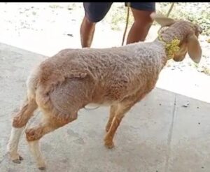
Fig 1. Cyst in thigh region
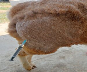
Fig 2. Fluid filled cyst
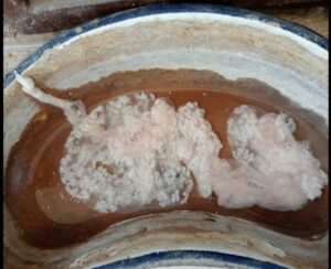
Fig 3. Coenurus cyst with scolices
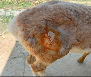
Fig 4. Post operative sutured skin
References:
Aiello, S.E. and Mays, A., The Merck Veterinary Manual, 8th edn. Merck & Co Inc. New Jersey, pp 320–324, 338–339 (1998).
El-Sinnary KA, Tageldin MH, Al-Sumry HS (1999) Outbreak of coenurosis (Taenia species) in anglonubian goats in the Sultanate of Oman. Vet Rec 144:296–297
Patro DN, Suhani B, Sahoo DK, Nanda SK, Pradhan PK, Nayak BC (1997) Incidence of generalised Coenurus gaigeri infection in a goat farm. Indian Vet J 74:68–69
Sharma DK, Singh N, Tiwari HA (1998) Prevalence and pathology of coenurosis in organized goat farms. J Vet Parasitol 12:30–32
Sharma DK, Chauhan PPS (2006) Coenurosis status in Afro-Asian region: a review. Small Ruminant Res 64:197–202a
Varma TK, Malviya HC (1989) Prevalence of coenuriosis in sheep, goat and pig in Bareilly, Uttar Pradesh. J Vet Parasitol 3:69