


MANAGEMENT OF THELITIS IN GRADED MURRAH BUFFALO
Ankit Kumar 1, Dharya Yadav 2
1Internee student, Veterinary College and Research Institute Namakkal, TANUVAS
2Third year student, Veterinary College and Research Institute Namakkal, TANUVAS
E-mail ID: ankitaiims357@gmail.com
Mobile no.: 8292265530
Abstract
Mammillitis is a condition in she buffaloes in which there will be sudden teat inflammation, enlargement, difficulty in milking, difficult for the calf to suckle, formation of black necrotic patches with circumscribed necrotic areas on teat base. The affected teat slough-off in primiparous buffaloes and the cause is suspected to be by Herpes virus. Vesicle formation, a characteristic feature of early stages of the disease in cow is either absent or occurs rarely in dairy buffaloes. Stress of calving and hormonal changes close to calving are important predisposing factors. It is more prevalent in buffaloes in their first lactation. Therapeutic interventions be used for the treatment of topical and parenteral administration of antibiotics, topical and oral administration of acyclovir (An anti-herpes virus drug), oral administration of zinc sulphate and other immunity enhancing agents and parenteral administration of antihistamine, lithium antimony thiomalate and anti-inflammatory drugs. Buffalo were treated with lithium antimony thiomalate (2 mg / kg deep IM), chlorpheniramine maleate (0.5 – 1.0 mg / kg I.M.), meloxicam (0.5-1.0 mg/ kg I.M.) and topically ethnoveterinary application. Clinical recovery was seen in buffalo after 6 days of last dose of treatment. The hyperemic and edematous teats noticed earlier before treatment slowly resolved to almost normal size.
Keywords: Buffalo, Mammillitis, Lithium antimony thiomalate, Acyclovir; Chlorpheniramine maleate, Meloxicam .
Introduction
Bovine herpesvirus 2 (BHV-2) causes mammillitis of teat in cows. There will be sudden onset of inflammation, papules, skin sloughing, vesicular growth, scab forming, and ulceration. In early lactating primiparous buffaloes, there will be an acute inflammation of one or more teats leading to necrotic ring formation, gangrene and fibrosis (Mouli, 1992 [14]). The condition is less successfully treated with antibiotics, NSAIDs and glucocorticoids. Lithium antimony thiomalate was used for the condition with better cure rate in early conditions. The probable mechanism of lithium antimony thiomalate in curing the condition might be due to its anti-fibrosis activity (Shridhar et al., 2014 [25]. The incidence of obstructive thelitis is noticed most commonly in first lactation buffaloes predominantly in graded Murrah than nondescriptive animals.
Examination of the milk sample
The milk from the affected quarter was subjected to California Mastitis Test (CMT) and further culture and sensitivity test (Christie and Koegh, 1940 [4]). The pH of the milk samples was also recorded. The somatic cell count of milk samples was did as per the method of Schalm et al. (1971 [19]). Electrical conductivity was measured using conductivity meter (Sneath Peter et al., 1986) [26].
Clinical Signs
The clinical condition has a wide range of symptoms. Without any physical abnormalities, some affected animals may become hard milkers. Others may have minor teat lesions, while a small percentage of those infected suffer severe teat lesions. (Abd-El-Hady, 2015; Sandrucci et al., 2014). According to (Sandrucci et al., 2014; Shearer et al., 2008), BHM in cows starts with painful raised edematous swellings of teats followed by appearance of vesicles (0.5 to 5 cm in diameter) which are irregular in shape. Within 24 h, these vesicles rapture leading to formation of ulcers that exude copious serum. Upon drying of this exudate, thick dark reddish-brown scabs appear on the teats. Healing usually occurs in 3 weeks, although ulcerated lesions may persist for months (Sandrucci et al., 2014). Teat and udder lesions may fuse together and may extend to the perineum resulting in vulvovaginitis. Milking causes pain to the affected animal and sometimes the entire teat becomes necrotic. Calves suckling affected cows may develop lesions in mouth (George et al., 2008). According to (Sharma et al., 1998), in dairy buffaloes, the lesions of BHM mostly occurred at the udder-teat junction of hind teats. The initial phase of the disease was characterized by the formation of localized plaques (2 to 5 mm) within the thickness of teat wall. These plaques were surrounded by inflammatory zones and ruptured within 48 h leaving very deep ulcers. Vesicle formation was not observed. As the healing progressed, a very thick blue-black scab developed and covered the healing lesion. Systemic signs of illness (e.g. rise in body temperature, pulse and respiration rates) were not present in the affected buffaloes. Milk of affected teats is usually negative for mastitis (Purohit et al., 2014). According to (Rao et al., 2003), in buffaloes, the size of the affected teat increases 2 to 3 times its normal size with a glossy appearance. Severe inflammation of teat causes tenderness and loss of flexibility. Thickening of the wall of the affected teat may result in narrowing of the teat canal and thus pain and difficulty in milking (Abd-El-Hady, 2015). In dairy buffalo, vesicle formation (a characteristics feature of early stages of BHM in cow) is either absent (Sharma et al., 1998; Sharma et al., 2005) or occurs rarely (Lokanadhamu et al., 2005). Annapurna et al., (2013) briefed about a case of fibroadenoma of the mammary gland in a buffalo heifer where they observed the fibro papillomma of the teat of the buffaloes.
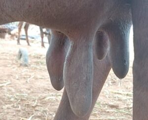
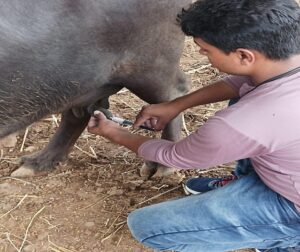
a)Photograph of graded Murrah buffalo b) Intramammary infusion of prednisolone
showing swollen and enlarged teat. and gentamicin.
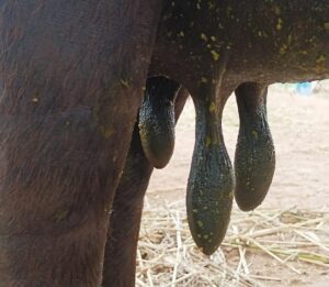
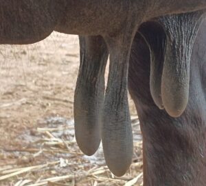
- c) Ethno-veterinary medicine topical d) Photograph of graded murrah showing
application. response to the treatment.
Treatment
Lithium antimony thiomalate @ 2 mg/kg deep IM, chlorpheniramine maleate 05-1.0 mg/kg IM, meloxicam 0.5-1 mg/kg, IM. Chlorpheniramine maleate is an antihistamine with known anti allergic property. Meloxicam is an NSAID with well-known anti-inflammatory activity (Shridhar et al., 2014 [25], Syed et al., 2009 [28]). Antibiotic (Enrofloxacin) were used to control secondary infection. EVM (ethno-veterinary medicine) application was done which includes aloe vera extract (250 gm), turmeric (50 gm), lime (10 gm).Intramammary infusion of prednisolone and gentamicin were also done. The treated buffaloes were kept under observation for 10-15 days and the recovery signs were observed and recorded.
Conclusion
Primiparous buffaloes in first calving do suffer from a condition known as mammillitis which in which there will be characteristic teat inflammation, enlargement, difficulty in milking, difficult for the calf to suckle followed by black necrotic ring formation and finally the teat will slough off from udder. The disease is caused by bovine herpes virus (BHV-2). The buffaloes suffering from the mammillitis were treated with lithium antimony thiomalate, acyclovir, chlorpheniramine maleate and meloxicam with good recovery rate which indicates the efficacy of these drugs in the therapy of buffalo mammillitis.
Reference
1.Kathirvel.S and Dharmaceelan.S 2016. Thelitis in Buffaloes – Reveiw of 12 Clinical Cases. Indian Vet. J., 93 (10): 73 – 74.
- Sharma, S, K B Singh, B.K. Bansal and D.K. Sharma,2005. Clinical symptomatology and epidemiological observations on teat skin lesions in buffaloes Buffalo Bulletin, 24(1): 12-16.
- Sreedevi, B., U.V.N.Malleshwar Rao and T. Venkat Reddy 2002. Proceeding of the IAAVR, Round Table Conference on Mastitis, No.3: 33-35
- Lokanadhamu M, Sreedevi B, Venkata Reddy T. Studies on etiology, symptomatology, diagnosis and therapy in ulcerative thelitis of buffaloes in Andhra Pradesh (India). Buffalo Bull. 2005; 24(3):56-69.
- Shridhar NB, Santhosh Kumar CN. Kadagi M.Therapeutic management of mammillitis- A case report of 5 buffaloes. Intas Polivet. 2014; 15(1):83-84.
- Kachawa, J. P., Ankita Sharma, Tanuj K Tanwar and Singh, A. P., 2017. Bovine ulcerative mammilitis and its therapeutic management. Ruminant Sci., 6 (1):193-194.