


OUTBREAK OF LUMPY SKIN DISEASE (LSD) IN CATTLE IN CHHOTANAGPUR PLATEAU REGION (INDIA)
Compiled, & shared by-DR. RAJESH KUMAR SINGH, (LIVESTOCK & POULTRY CONSULTANT), JAMSHEDPUR Post no 1380 Dt 31/08//2019
JHARKHAND,INDIA 9431309542, rajeshsinghvet@gmail.com
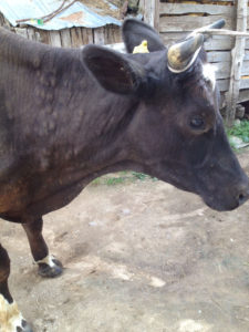
After the arrival of monsoon in India particularly in the Chhotanagpur plateue region which covers parts of Odissa, Jharkhand, WB and Chatisgarh, the level of humidity become very high during moist weather leading to sever fly menace.These flies bite the livestock which causes hypersensitivity of the skin lesions but some time also spread diseases like cow pox better to say lumpy skin disease among the animals.There has been reports from field veterinarians of Odissa, Jharkhand,WB regarding the outbreak of this LSD in cattle.Here in this post I have tried to through a brief light on this on going outbreak.
Lumpy skin disease virus causes a severe disease in cattle characterised by nodules in the skin. Transmission of LSD occurs via insect vectors and vaccination is the most effective means of control. During the past five years lumpy skin disease has spread through the Middle East into southeast Europe, the Caucasus, southwest Russia and western Asia. The disease causes substantial losses in affected herds with significant economic consequences. It also blocks access of affected countries to lucrative export markets, compounding the financial impact of a LSD outbreak. The main lesson to be learnt from the current European LSD epidemic is to be vigilant of emerging diseases.
Preferred Scientific Name
• lumpy skin disease
International Common Names
• English: lumpy skin disease, capripoxvirus, in cattle- exotic; yak pox
• French: dermatose nodulaire contagieuse; la dermatose nodulaire contagieuse
English acronym
• LSD
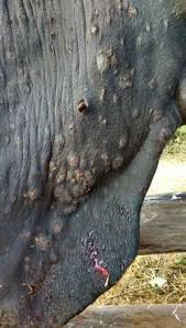
Lumpy skin disease (LSD) is a pox viral disease of cattle with a major socio-economic impact. The disease is characterized by fever, multiple firm, circumscribed skin nodules, and necrotic plaques in the mucous membranes (chiefly of the upper respiratory tract and oral cavity), mastitis, orchitis and swelling of the peripheral lymph nodes. The disease is caused by a capripox virus of which the prototype strain, “Neethling’” was first isolated in South Africa. Clinically, the skin lesions of LSD closely resemble those of pseudo-lumpy skin disease caused by the Allerton strain of bovid herpesvirus 2 (BHV-2).
Lumpy Skin disease (LSD) is a condition caused by a capripox virus of the family Poxiviridae, which is diagnostically indistinguishable from sheep pox virus. It affects cattle only and sheep and goats do not become infected during outbreaks of LSD even when held in close contact with infected cattle. The capripox virus strains are identical in every way in the laboratory, differing only in their ability to produce LSD in one host and sheep and goat pox in the other ,The clinical syndrome of lumpy skin disease was first described in Zambia in 1929. It was at first, considered to be the result of hypersensitivity to insect bites or poisoning. The disease is now widespread across Asian countries and is enzootic in sub-Saharan Africa.
Transmission is mostly via insect vectors, although direct contact can transmit the virus. Pox viruses are highly resistant and may remain viable in infected tissue for at least four months, and probably longer. Virus is also present in blood, nasal and lachrymal secretions, semen and saliva, which may be sources for transmission. It is not known if a particular insect is the main vector, as the virus has been found in the Stomoxys spp., and the Biomyia fasciata mosquito species. Tabanidae, Glossina and Culicoides spp. have all been found in situations where there has been ongoing LSD transmission and have been suspected to be involved. Considerable research into the transmission of the disease still needs to be undertaken in order to fully understand the condition and devise control measures for it.
Mortality rates in this condition can reach around 40% and morbidity rates are considerably higher. Skin lesions result in severe and permanent damage to hides, which is a cause of economic loss, while lesions in the mouth, pharynx and respiratory tract cause a rapid decrease in body condition, which may persist for several months.
Classification
OIE List A disease
Transmission
Capripox viruses are very resistant and remain viable for long periods, on or off the animal host e.g. they may persist for up to 6 months in shaded animal pens, and for at least 3 months in dry scabs on the fleece, skin and hair from infected animals.
Transmission of SGP is by contact with infected animals, their aerosols, nasal secretions, saliva or dried scabs. The viruses are also readily transported on clothes and equipment. Insects e.g. the stable fly (Stomoxys calcitrans) act sometimes as mechanical vectors.
No specific information is available on transmission of SGP through semen or embryos.
NB With lumpy skin disease in cattle, virus has been detected in semen for up to three weeks after infection.
Risk of introduction
Movement of infected animal is the main means by which SGP is spread to new areas. Thus where there are no importations of live animals or genetic material from endemically infected countries there is little risk.
However, the viruses can be transported on clothing, equipment and unprocessed wool products brought in by people from affected countries. Therefore the role of quarantine is of primary importance.
Signalment
Cattle of all ages can be infected by the virus. There is no sex predilection. Channel Island breeds are much more susceptible to the virus.
Clinical signs
The diseases are more severe in lambs and kids than adults. Disease begins with:
• Sudden onset of fever
• Discharges from the nose and eyes, and salivation
• Loss op appetite
• Reluctance to move
• Skin lesions appear in 1-2 days, extending all over the skin, but are most obvious on face, eyelids and ears, perineum and tail. Lesions may also be seen on the mucous membranes of the nostrils, mouth and vulva
• Acute respiratory distress
• Mortality peaks about two weeks after the onset of the skin
lesions
Lesions begin as an area of reddening, progressing over two weeks to a papule, vesicle, pustule with exudation and scab formation. Healing is very slow.
A nodular form of the disease (‘stonepox’) can also occur this resembles lumpy skin disease in cattle.
Post-mortem findings
The epidermal and mucosal lesions described above will be apparent at post-mortem.
Ulcerations may be found in the lining of the trachea and gastro-intestinal tract. Lung lesions consisting of pale grey nodules may also be seen.
Differential diagnosis
• Contagious pustular dermatitis (scabby mouth)
• Bluetongue
• Mycotic dermatitis
• Sheep scab
• Mange
• Photosensitisation
Specimens required for diagnosis
SGP should be suspected when an acute disease of sheep or goats is accompanied by fever, pox-like skin lesions and high mortality.
For serological examination, paired of blood sample from animals with fever.
For virus identification, samples can be sera, vesicular fluid, scabs and skin scrapings of lesions and lesions in the respiratory and gastro-intestinal tract. Samples should be taken within the first week of apparition of the symptom and be kept fresh. OIE recommends to use the identification methods which could be cell inoculation and identification by immunofluorescence, staining of intracytoplasmic inclusion bodies, inhibition of cytopathic effect using positive serum or ELISA.
Signs include pyrexia of 40 – 42oC, anorexia, depression, lethargy and excess lacrimation. Soon after this onset, dermatological signs begin to develop which appear as round circumscribed areas of erect hair measuring between 5 to 50mm in diameter. These lesions are raised and firm and they may be surrounded by a ring of haemorrhage. The regional superficial lymph nodes are enlarged and oedematous.
Other signs include nasal discharge and ptyalism, which is thought to be due to lesions in the nose and mouth. Lesions can be found in the respiratory tract and alimentary tract and so can cause coughing, increased respiratory noises and diarrhoea. The lesions are often secondarily infected by bacteria causing any discharge to be purulent and pneumonia is a common sequelae of the disease. Lesions may eventually slough away to leave a hole of full skin thickness, known as “sitfast”.
Clinical signs and pathology———-
A morbidity rate of five to 45 per cent on affected farms is usual. The mortality rate may be as high as ten per cent, even among indigenous cattle. Typically, cattle develop a biphasic febrile response two to four weeks after exposure to the virus. Animals remain febrile for four to fourteen days, during which time they may develop salivation, lachrymation and a mucoid or mucopurulent nasal discharge. Lachrymation may be followed by conjunctivitis, corneal opacity and blindness in some cases. In the majority of animals the superficial lymph nodes are enlarged. Skin nodules, the characteristic feature of the disease, appear before or during the second rise in body temperature, four to ten days after the initial febrile response. The nodules, which are randomly distributed and range in diameter from 10 to 20 mm, involve both the skin and subcutaneous tissues and sometimes even the underlying musculature. The size of the nodules is usually fairly uniform but several nodules may fuse to form large, irregularly circumscribed plaques. The number of nodules may range from a few to several thousand in severely affected animals. The nodules are well-circumscribed, firm, round and raised and are particularly conspicuous in short-haired animals. In long-haired cattle the nodules are often only recognized when the skin is palpated or moistened. In most cases the nodules are particularly noticeable in the perineum and on the vulva. Skin lesions resolve, become indurated (in which case they persist as hard lumps or “sitfasts” for 12 months or longer) or sequestrated to leave deep ulcers partly filled with granulation tissue that often suppurates. In severely acutely affected animals the ventral parts of the body, for example the dewlap and the legs may be slightly oedematous one to two days before the appearance of the nodules. In these acute cases the oedema of particularly the legs may become very severe following the development of the nodules. Nodular skin lesions may extend into underlying tissue such as tendons and tendon sheaths resulting in lameness in one or more legs. Most affected animals have multifocal, roughly circular, necrotic areas on the muzzle and in the respiratory tract (nasal cavity, larynx, trachea and bronchi), and buccal cavity (the inside of the lips, gingivae and dental pad), but these lesions may also be present in the fore stomachs, abomasum, uterus, vagina, teats, udder, and testes. Generalized lymphadenopathy comprising lymphoid hyperplasia and oedema is a regular finding.
Diagnosis
In countries where the disease is endemic, diagnosis based on clinical signs is generally all that is required as veterinarians here are experienced at detecting the lesions. In countries where the disease is exotic, signs may be confused with other diseases such as bovine herpes virus 2, foot and mouth disease, insect bite hypersensitivity or demodicosis.
Samples should be taken from the lesions and can be viewed under electron microscope to identify pox-virus particles.
Samples of skin also show characteristic histopathological changes which include vasculitis and perivascular infiltration with white cells causing a thrombosis of the vessel in the dermis and subcutis. Cells infiltrating the lesion are epithelial cells, known as celles clavelauses, which are also described in sheep pox.
Virus isolation by culture can also be carried out, but this takes many days and is not useful diagnostically for this reason.
Treatment and Control
Treatment is supportive, in the form of wound dressing to prevent fly strike and secondary infections. If secondary infections have already occurred, systemic antibiotics can be given to treat these accordingly.
In countries where the disease is exotic, measures to control the disease would include immediate slaughter of all infected animals and in-contact animals and the carcasses destroyed. A vaccination cover with a 25 to 50 km radius around the focus may then be established and all cattle movements stopped within that zone, however, this vaccination policy may allow the virus to persist in some cattle. Where an epizootic infection occurs in an already enzootic area, slaughter policies are inappropriate and vaccination campaigns are recommended instead. Vaccination will greatly reduce the morbidity and economic effects of an epizootic infection. Follow-up vaccination of calves and re-vaccination programmes over a period-of two to three years will greatly reduce the incidence of clinical disease. No country in sub-Saharan Africa, however, has succeeded in eradicating LSD once it has occurred. Movement should be restricted to prevent new spread of the disease in these situations also.
Two different vaccines have been widely and successfully used for the prevention of LSD in cattle populations in India. The Neethling strain vaccine has been used has proven effective in reducing the clinical signs of disease in cattle. Some local reactions do occur in many animals following inoculation, but these are not severe.
During tis outbreak in our area we are giving parental therapy of antibiotics, antihistaminic,anti inflamatory , multivitamins and topical use of ointment.Besides this, the farmers are advised to wash the skin lesions with neem and karanj oil.
Control / vaccines
In endemic areas systematic vaccination programs have provided the most effective control over the disease.
In an outbreak situation diseased sheep or goats should be destroyed and their carcases burnt or buried. This could be supplemented by vaccination of in-contact animals, and movement controls for both animals and vehicle should be applied.
Cell-cultured attenuated and inactivated vaccines have been used to prevent disease. Inactivated vaccines provide about five months protection but there is no ready commercial source. The live attenuated vaccines give good immunity and are considered suitable in an emergency situation.