


PHYSIOLOGY & ROLE OF THE SKELETON BONE & CALCIUM IN EGG PRODUCTION IN LAYING HENS
The skeletal system provides the strong framework for the support and protection of the remainder of the systems, organs and tissues that make up the body of the fowl. Bird bones that are homologous to bones found in other animals have evolved over time to enhance the ability of the bird to fly. While fowls are not able to fly well, they still retain that ability to some extent. These modifications include:
- Pneumatic bones where the air sacs of the respiratory system connect with the cavity of some of the long bones.
- Fusion of some vertebral sections to provide the rigidity required for flight.
- The sternum provides a large surface area for the strong attachment of the main muscles used for flight.
- Compared to other animals, the size of the head has been reduced significantly as a large head would be a hindrance when flying.
- The neck is quite long in most bird species to enable:
- Protection of the delicate tissues of the brain from too much jarring when landing. The flexibility of the neck acts as a shock absorber.
- The bird to reach food located on the ground – a rigid body makes this simple activity more difficult.
- The bird to adjust its centre of gravity when the bird changes from the upright position of walking or perching to the more horizontal position of flight.
- The long tail of many other animals has been reduced to a very short section of fused bones called the pygostyle.
- The ribs have been modified by the inclusion of the uncinate process – a rearward projection of bone – which gives strength to the rib cage.
The anatomy and physiology of birds show specific adaptations for the egg laying. Laying hens have a unique calcium and bone metabolism, adapted for egg production and eggshell formation.
Because of productive, economic and animal welfare reasons, it is essential to understand the structure and function of the bone and the calcium metabolism in laying hens. This knowledge will help us to apply the correct management and feeding strategies in order to achieve appropriate mineral reserves in the skeletal system, an eggshell of good quality, and the prevention of problems or imbalances derived from an alteration of these factors.
It is worth mentioning that, in addition to the skeletal system, the digestive system, including the liver and the intestine, is key for laying hens.
The avian skeleton is a unique system that is specialized for flying, walking on two legs and laying eggs. Establishing and maintaining a strong skeleton is vital to ensure a productive laying hen. In order to understand the impact of diet on the laying hen, it is important to understand the biology of the skeleton.
There are three different types of bone: cortical, trabecular and medullary.
- Cortical bone is the hard outer surface of the round bones, such as the femur or the humerus, and the fl at bones, such as the skull or the pelvis.
- Trabecular or spongy bone is less dense than cortical bone and helps support the structure inside the cortical bones.
- Medullary bone is a specialized woven bone which serves as a calcium reserve for the demands of egg shell formation. Easily created and resorbed, medullary bone is ideally the first source mobilised when more calcium is required.
While the outside appearance of avian bones is similar to those of mammals, there are several key differences.
- Fused vertebrae – Several thoracic and lumbar vertebral spinal sections are fused together to form a more solid structure for flying.
- Keel – The sternum or keel provides a large surface for attachment of the pectoral muscles, which are important for energy storage and muscle yield.
- Pneumatic bones – Hollow and air-filled, these bones are part of the respiratory system and help with flying.
- Medullary bone – This specialised bone is used as a source of calcium for the egg shell and only occurs in birds and some reptiles.
Bone growth and resorption is controlled and regulated by a few important cells and many different hormones. In healthy, well-fed birds, the cells and hormones work closely together to maintain bone structure and blood calcium levels needed for optimum production.
The important cells for bone growth and modelling are chondrocytes, osteoblasts, and osteoclasts.
Chondrocytes start the basic process for bone growth by secreting type II collagen and other important components for bone formation.
Osteoblasts then produce the type I collagen and the increased levels of calcium and phosphate that result in the mineralisation or ossification of the bone.
Osteoclasts resorb bone for remodelling or for releasing calcium into the blood stream. Bone growth and resorption is regulated by a number of different hormones which control when structural or medullary bone grows or resorbs, depending on the physiologic need.
Growth hormone stimulates cellular growth and protein synthesis throughout the body.
Thyroxine stimulates cell metabolism as well as osteoblast activity.
Melatonin influences osteoblast activity. Melatonin levels are highest when birds are sleeping during the dark period and initiate a cascade of events affecting hormones necessary for egg production.
Oestrogen increases at sexual maturity and changes osteoblast activity from creating cortical and trabecular bone to creating medullary bone. After the first egg, the only way a hen can remodel structural bone is during periods of low oestrogen, such as moult or breaks in lay during the normal production period.
Calcitonin is released when there are high serum calcium levels and decreases osteoclast activity while increasing osteoblast activity which builds bone and lowers serum calcium levels.
Parathyroid hormone (PTH) is released during periods of low serum calcium and binds to osteoblasts. This binding decreases osteoblast activity while releasing a compound that increases osteoclast activity, thus increasing serum calcium levels. Additional properties of PTH include increasing small intestine absorption of calcium and decreasing urinary excretion of calcium.Calcitonin and parathyroid hormone work together in feedback loops to ensure the proper levels of serum calcium are maintained.
Bone formation
The skeleton provides support and protection for the remainder of the systems and tissue. Bone is living tissue and its structure is largely affected by the nature of stresses placed upon it. The chemical composition is also quite variable although it mainly consists of calcium and phosphorus in the form of hydroxyapetite [3Ca3(PO4)2.Ca(OH)2] crystals deposited on a fine matrix of collagen fibres, along with other cell types.
Deposition And Adsorption Of Bone
The metabolic activity in bones is continuous and the microscopic structure is constantly changing. Small cells, called osteoblasts, are responsible for depositing new bone tissue, while large polynucleate cells, called osteoclasts, resorb existing bone. Other cells, called osteocytes, found in the calcified mass of bone, help maintain the bone structure. Thus the skeleton is a major reservoir of calcium and phosphorus. Therefore, it is very important to maintain the proper levels of these minerals in the diet (99% of body calcium and 80% of body phosphorus are stored in the skeleton). Sodium and magnesium are other minerals of importance in bone structure. These may be drawn upon when the diet of the animal contains inadequate supplies.
Microstructure
The microstructure of the bone changes continuously as bone is a target for a number of influences. Chemicals, in particular calcium and phosphorus, are continuously added or removed from the bone. Other influences include:
- The level of certain hormones e.g. growth hormone, parathyroid hormone, calcitonin, oestrogenic and androgenic hormones in the blood
-
- The level of vitamin D in the diet
Young chickens are very sensitive to vitamin D deficiency. This vitamin is required by the chicken for the assimilation and use of calcium and any deficiency will be seen as a typical calcium deficiency, such as rickets. Vitamin D is found in a number of slightly different forms and cholecalciferol (D3) is ten times more active than ergocalciferol (D2) in preventing rickets.
Ergosterol, a compound under the skin of animals including poultry, is converted to usable Vitamin D by the rays of the sun. In the layer hen, the skeleton is particularly vulnerable to the demands made for calcium for eggshell formation.
Stages Of Bone Development
All bones of the body are formed in pre-existing tissues that they either replace or use in their structure. It is usual to find that bones pass through three stages as they develop:
- Prechondral or membranous stage
-
- Chondral or cartilaginous stage
- Ossification stage (bone formation)
Most of the bones of the fowl go through the cartilaginous stage. A few such as the bones of the skull omit this stage. As far as the chicken is concerned, the membranous stage takes place in the egg during embryo development. Only the cartilaginous stage and the ossification stages are easily identified.
The secretion of special cells called chondroblasts, brings about cartilage formation. The ossification process then hardens the cartilage when the bone takes up minerals, mainly calcium carbonate. Long bones increase in length by the ossification process. In birds much of the bone is laid down in successive layers to form dense, compact bone covered by the cellular periosteum on the outside. Long bones are usually hollow with the hollow filled with bone marrow and extensions of the air sacs.
Compact bone is modified by the formation of special cavities that eventually mineralise by depositing concentric layers of new bone. The new structure is called the haversian system. If a transverse section (slice) of bone is examined under a microscope, a large number of small canals that run more or less parallel to the long axis will be seen. These canals are called haversian canals and carry the blood vessels and nerve fibres. Surrounding these canals are plates of bone and between the plates are small spaces called lacunae. A special bone cell called an osteocyte is found in each lacunae. Nutrients pass from the blood vessels in the haversian canals to the bone through small canals called canaliculi.
Medullary Bone (Layer Fatigue)
A very unique feature of the female avian skeleton is the way the bird lays down a supply of highly unstable secondary bone in the marrow cavities of bones during the reproductive period. This bone is called medullary bone and because of its instability, provides a very ready source of calcium for eggshells. In a production hen, not enough calcium can be absorbed across the intestinal wall in one day supply to satisfy the requirements for production of an egg shell. Without this medullary bone the eggshells would be very thin and weak.
Medullary bone starts to develop about 10-14 days before the first egg is laid as a result of the presence of oestrogen and androgen in the blood as the pullet reaches sexual maturity. This function remains for the length of her laying life. Approximately two weeks before egg production starts the pullet flock should be changed from the growing to the layer diet which is higher in calcium. If pullets come into production at too young an age, they may deplete body reserves of calcium that may result in thinner shelled eggs and /or a drop in production. This should not occur if production is delayed to an age best suited to the strain of layer.
If a calcium deficient diet is provided to a layer, it will deplete her skeleton of calcium and thus make the bird significantly weaker. Eggshells will also become thinner and production will decline until it ceases altogether. A condition of paralysis, called cage layer fatigue, may be seen in layers housed in laying cages. When seen, it is always associated with birds in very high production and takes the form of muscular paralysis and osteoporosis (weak bones). While the cause is not fully understood, the birds usually recover quite quickly when taken out of the cages and placed on the floor for a short period. The weaker shells of eggs from older hens are caused by a loss in efficiency by the oviduct shell glands in their production of the shells.
- SOURCES OF CALCIUM: FEED AND SKELETON
The high levels of calcium required for the eggshell formation are obtained from different sources: the diet (through intestinal absorption) and the body (through bone resorption and reduced renal excretion of calcium). The control of the calcium levels in the plasma is done through hormonal regulation, which maintains the balance between these three mechanisms of calcium obtention.
The calcium absorption capacity at the intestinal level is an important quality in high-production layers, since this factor should guarantee the calcium supply for the formation of the eggshell and its thickness, as well as its capacity to withstand handling without breaking.
When the calcium from the diet is insufficient, or it is not absorbed correctly, the bird obtains the calcium from the bone reserves through a process called bone resorption, defined in the following sections.
- ROLE OF THE BONE IN THE LAYING PERIOD
- ANATOMY AND PHYISIOLOGY OF THE BONE
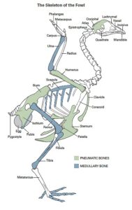
The skeletal system of birds is lightweight to facilitate the flight, thanks to the fusion of several bones into one and the large presence of pneumatic bones, which contain air instead of bone marrow. The bones with bone marrow are the long bones distal to the humerus and pelvis (Image 1).
Bones are made up of a collagen matrix that surrounds the cellular component, made up of osteoclasts, osteocytes, and osteoblasts. The different types of bone cells and tissues, as well as the bone metabolism, are described below.
Types of bone cells
Image 1: Classification of the types of bones od the skeleton of the hen. (Source: Hy-Line, Technical Bulletin – Understanding the role of the skeleton in egg production)
The bone-creating action of the osteoblasts, together with the bone destruction of the osteoclasts, forms the process of bone remodeling, intended to preserve the mechanical integrity of the skeleton.Osteoblasts are bone-forming cells. When these cells are active, they secrete proteins and alkaline phosphatase for the synthesis of new bone matrix and its mineralization. During bone formation, some osteoblasts remain in the matrix they deposit, and differentiate into osteocytes, with structural function. Osteoclasts are responsible for bone resorption by releasing hydrogen ions, that dissolve the mineral phase of bone and proteolytic enzymes that degrade collagen.
Types of bone tissue
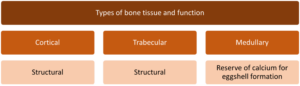
There are three different types of bone tissue: cortical, trabecular, and medullary bone.
The cortical bone is tubular in shape to enclose the medullary cavity, and makes up the outer structure of round bones, providing great strength to the bone.
The trabecular bone, located in the inner part of the cortical bone, is less dense than the first one and has a faster and more efficient osteogenic surface. Thus, it experiences greater remodeling. These two types of bones are present in all birds and have a structural function to support the skeleton.
The medullary bone is characteristic only of laying hens. It is developed during the sexual maturity under the effect of estrogens, and it is easily mobilized. Because of that, it acts as a reserve of calcium for the eggshell formation when calcium from the diet is insufficient.
Bone metabolism
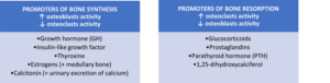
Bone formation and resorption are regulated by several cells, the osteoblasts and osteoclasts, mentioned above, and different hormones, which are named with their main activity in the bone in the following table :
Estrogens decrease bone resorption. Therefore, bone remodeling occurs when the estrogen levels are low, like in the moulting or during daily rest in posture.
This article highlights the process of calcium release from the bone and its hormonal regulation by calcitonin and parathyroid hormone (Image 2):
- Calcitonin is a hypocalcemic hormone that inhibits bone resorption. In addition, it acts on the kidney, increasing the urinary excretion of calcium to reduce serum calcium levels.
- Parathyroid hormone (PTH) has the opposite action to calcitonin. It is hypercalcemic and it is released during periods of low serum calcium. PTH has acts directly on the bone and the kidneys, and indirectly on the intestine. It binds to osteoblasts, decreasing their activity, and activates osteoclasts, promoting bone resorption. At the same time, it stimulates the reabsorption of calcium in the urine, which is exchanged with the elimination of phosphorus. At an intestinal level, it acts on the mucosa of the small intestine, increasing the absorption of calcium from the diet.
- DEVELOPMENT OF THE SKELETON IN PULLETS AND SEXUAL MATURITY
The development of the intestinal tract occurs, mainly, during the first weeks of life of the pullets and is vital for the absorption of nutrients and the productive efficiency of the future layer. During this time the skeleton is also under development.
The highest growth rate of the structural bone occurs between weeks 6 and 12. At the end of this period, the size of the bird is established, since 95% of the skeleton is developed, although it has only 75% of its mature weight. Any delay in growth will affect the size of the adult bird and delay the start of production.
Weight gain in the later 6 weeks after this phase will correspond to the development of the muscle, the reproductive tract, and the medullary bone, which will be completed by week 32 of age.
When the animal is close to the sexual maturity, there is an increase in estrogen that closes the growth plates and stops the development of structural bone. In turn, this hormone stimulates the formation of medullary bone on the inner surface of the structural bone.
- BONE REMODELING PROCESS LINKED TO THE LAYING PERIOD
Bone content and mineral density, as well as the proportion of the different types of bone (cortical, trabecular, or medullary) can change dramatically during the laying period.
This is because both osteoclastic activity (bone resorption) and osteoblastic activity (bone formation) are present simultaneously during the ovulatory cycle and will modify the structure of the bone. Depending on the calcium intake rate and the hormonal activity, one will predominate over the other.
Eggshell formation takes place, usually, during darkness and after several hours since the last feed intake. By that time, the serum calcium levels are very basic, and the bone is responsible for the calcium supply thanks to the osteoclastic activity, mainly from the medullary bone. After oviposition, there is a short period of rest until a new eggshell begins to form, which is the time when the hen redeposits the medullary bone.
- PROBLEMS DERIVED FROM THE REDEPOSITION OF THE MEDULLARY BONE
Immature birds have a thick cortical bone and a good trabecular structure on the inside, but they lack medullary bone. As sexual maturity arrives, the medullary bone is deposited on the inner surfaces of the trabecular and cortical bone, which protects the structural tissues from bone resorption.
Over time, hypocalcemic periods when the medullary bone is mobilized cause a gradual diffusion of the medullary bone through the spaces of the structural bone, when it is redeposited. This diffuse deposition does not provide the same level of protection over structural bone to prevent its resorption and, therefore, mobilization of both trabecular and cortical bone occurs.
At the end of the production cycle, the cortical bone layer is very thin, few trabecular structures remain, and the medullary bone is widespread in the medullary cavity.
Because of this, the problems of fractures, crooked keels, osteoporosis, or cage fatigue syndrome that may appear towards the end of the laying period can be related to the stability of the structural bone, rather than with the reserve of medullary bone.
- ORIGIN OF THE LOSS OF INTEGRITY OF THE SKELETON OF LAYING HENS
Nutritional deficiencies are usually the first cause of the decrease in the integrity of the hen’s skeleton that may lead to problems in the eggshell quality.
The low mineral intake, especially of calcium, due to insufficient supply or availability; intestinal dysbiosis affecting intestinal integrity and absorption; as well as feed restriction during the dark period, which is the moment when eggshell calcification is greater, lead to a predominant use of calcium of bone origin that can end up destabilizing the structural bone and generate problems of prostration, fractures, cage fatigue syndrome and an increase in the rate of broken eggs, consequences which have already been described in the previous section.
Heat stress is another important factor that causes the loss of the reserves of bone calcium. When animals hyperventilate, respiratory acidosis occurs. This is compensated by the mobilization of carbonates from the bone. These carbonates are bound to calcium, so there is a loss of this mineral, and, indirectly, the quality of the eggshell is affected.
- EGG SHELL FORMATION
The eggshell is made up of approximately 95% calcium carbonate, mainly in the form of calcite crystals, and 5% of organic material, in the form of membranes and an organic matrix.
The eggshell formation process consists of different stages. Initially, the deposition of water, salts and glucose occurs, which increases the volume of the egg and works as a stimulus to initiate the rapid calcification of the eggshell.
The maximum rate of calcium deposition occurs between 12 and 18 hours after ovulation, when the egg is in the uterus. The calcium transfer from the blood to the eggshell at that time is very high (rates of 100-200 mg/h).
- STRATEGIES TO IMPROVE BONE QUALITY AND AVOID EGGSHELL PROBLEMS
Biovet S.A. has developed Alquerfeed Layers to reduce the use of calcium of bone origin, avoid the weakness of structural bone and the related problems in the eggshell quality, and maximize intestinal absorption of calcium for egg production. Alquerfeed Layers is an oral solution based on the combination of:
- Minerals as an additional calcium source to that of the diet, crucial in periods of high demand.
- Carbonates to prevent the loss of calcium reserves in situations of heat stress.
- Pronutrients, compounds of plant origin that maximize calcium absorption capacity at intestinal level.
In this way, the product aims to maintain the internal balance of the birds and optimize the laying process in order to extend the laying peak, slow down the post-peak decrease in production, and prevent egg breakage and fractures or the cage fatigue syndrome.
It is unlikely that there is another animal that can consume, absorb, transport and metabolize more calcium per unit of weight than birds.
Bone tissue is physiologically active and plays an important role in the homeostasis of serum calcium levels to compensate the temporary lack of intestinal calcium using the bone for the eggshell formation.
Consequently, the continuous bone remodeling that occurs due to the use of calcium from the bone, as well as the loss of calcium derived from heat stress situations, can weaken the skeleton of the bird and lead to bone disorders, such as fractures or cage fatigue syndrome, and decrease the eggshell quality, increasing the rate of broken eggs.

ROLE OF THE SKELETON IN EGG PRODUCTION
Compiled & Shared by- Team, LITD (Livestock Institute of Training & Development)
Image-Courtesy-Google
Reference-On Request.
SOURCE-https://www.hyline.com
HYLINE & https://www.thepoultrysite.com/