


POST MORTEM (PM) OR NECROPSY FINDINGS IN MAJOR POULTRY DISEASES
Post mortem inspection covers the inspection of the carcasses and parts of meat and poultry used for human food. It takes place after the poultry has been slaughtered or dies due to any infection. Thus, the term ‘post mortem’, which means ‘after death’ in Latin is used. The purpose of post mortem inspection is to protect the public health by ensuring that the carcasses and parts that enter commerce are wholesome, not adulterated, properly marked, labelled and packaged. It is also a very important diagnostic tool that is used to support other procedures performed in the diagnosis of disease conditions in birds. There are several other reasons for performing post mortem examination which includes finding the cause of death, confirming a diagnosis, investigating unsuccessful therapy, increasing knowledge or satisfying curiosity. Necropsy procedures are performed in a systematic manner. This is performed at regular basis in organized poultry farms to check the mortality rate. The procedures include collection of history, external examination and preparation of carcass, opening the body cavities, examination of organs and finally the carcass should be well disposed after proper examination to avoid spread of disease to human and animals.
Using a knife or scissors, a person can perform a basic necropsy to obtain diagnostic information, samples for further laboratory testing, or to ensure quality control of a flock. If you see a rise in mortality (death rates) or a rise in morbidity (number of ill birds), the necropsy can provide you with more information about the disease, perhaps even a diagnosis.
Basic Necropsy Needs
- A flat hard surface in a well-lighted area.
- Access to water and towels.
- Knife or scissors.
Consider gloves and a face mask if you suspect a potentially zoonotic disease (transmissible to humans) as the cause of illness or death.
Performing a Necropsy
- Wet down the feathers with a disinfectant solution to limit the distribution of feathers during the dissection.
- Place the bird on its back with its feet towards you.
- Grasp both legs and push down and away from the pelvis to loosen the joints.
- Tent the skin over the abdomen and cut with scissors or knife.
- Remove the skin overlying the abdomen and breast (from neck to cloaca).
- Examine the breast muscle for decreased muscle mass, paleness (anemia), or bruising.
- Incise the abdominal muscle and cut through the ribs on the sides of the keel bone.
- Grasp the keel near the abdomen and pull upwards to expose the internal organs and chest cavity.
- Examine the liver for changes in size or discoloration, white or yellow spots, abscesses, and/or tumors.
- Examine the air sacs for increased thickness and increased cloudiness. The normal air sac surfaces look like soap bubbles or clear cellophane wrap.
- Cut the gastrointestinal (GI) tract between the esophagus and proventriculus.
- Remove the proventriculus, ventriculus (gizzard), small intestines, large intestine, ceca, and cut off at the level of the cloaca. The pancreas will also be removed. It is the pinkish tan organ cradled within the loop of duodenum (a section of the small intestine).
- Cut all attachments close to the intestines and set the GI tract aside. At the end of the necropsy, these organs can be opened up and examined for internal parasites.
- Next, remove the liver and spleen. A green discoloration of the liver near the gall bladder is a normal finding. The spleen is the reddish, round organ located at the junction of the proventriculus and gizzard.
- Now you can observe the organs located near the backbone of the carcass.
- Examine the kidneys, which are elongated, lobulated organs that are embedded in the backbone of the bird, and the left ovary/oviduct (or paired testes), which are positioned on top of the kidneys.
- The lungs, which are attached to the ribs, can be gently teased out of the ribcage for further examination.
- The outer surface of the heart should be examined for a cloudy, thickened appearance, suggesting pericarditis. Also, note if excessive fluid is located between the heart and the pericardium (membranous covering of the heart).
- Next, turn the bird around to face you and cut through the corner of the beak.
- Extend the cut through the throat and down towards the heart.
- Examine the interior surface of the esophagus and crop. Look for the presence of food and/or parasites (worms) in the crop. If the inside surface appears to resemble a towel, it may be an indication of a fungal infection called “crop mycosis.”
- Next, cut through the larynx, trachea, and syrinx. The inside surface should be free of excess mucus.
- Turn the bird back to the previous positioning — feet in front of you.
- The sciatic nerve located on the interior upper thigh (located under muscle) should be exposed on both legs. The nerves should be the same size bilaterally with no swellings. Enlargement of this nerve can be an indication of Marek’s disease.
- With a sharp knife, cut through the stifle and hock joints, looking for yellow or white pus-like material, blood, or excess fluid. Joints should appear shiny and white with just a small amount of clear, sticky fluid inside.
- To find the bursa of Fabricius, cut through the cloaca and look for a grape-like structure towards the rear of the bird. The older the bird—the smaller the bursa. The bursa diminishes in size as the bird reaches sexual maturity.
- Cut the bursa in half. It should have wrinkles running parallel to each other on the surface and be cream colored in appearance. Note any discoloration or swelling.
- Now return to the GI tract and starting with the proventriculus, cut lengthwise. The inside wall is bumpy and this is normal as these are digestive glands.
- Cut through the ventriculus, intestines, and ceca. Note the appearance of the inside walls (mucosa) and the presence of parasites (worms), blood, and/or a thickened or discolored surfaces.
- Dispose of the carcass properly and disinfect surfaces and tools.
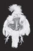
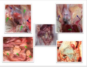
Figure A. The skin overlying the breast and abdomen is removed. The arrowhead is pointing to an abnormally crooked keel.
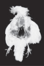
Fig. B. The keel is lifted and the liver is visualized. The normal liver should not extend beyond the tip of the keel.
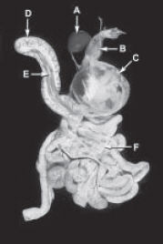
Figure C. The GI tract has been excised and positioned for further examination of the digestive organs. A = spleen. B = proventriculus. C = ventriculus. D = duodenum. E = pancreas. F = jejunum.
Post mortem findings in different diseases in poultry birds
Infectious bursal disease
It is an acute, highly infectious lymphocidal disease of young, sexually immature chickens caused by virus belonging to Birnaviridae family. In post mortem, we notice haemorrhagic and atrophied bursa with presence of white caseated material in the lumen of bursa, haemorrhages in thigh and pectoral muscle, haemorrhages at the junction between proventriculus and gizzard. Kidneys appear swollen and pale with accumulation of urates in tubules; liver appears pale and bile stained with necrotic foci.
Avian influenza
It is caused by influenza virus belonging to family orthomyxoviridae. Post mortem findings include subcutaneous edema of head and neck; inflammation of sinuses, trachea, air sacs and conjunctiva; congestion of musculature and dehydration, haemorrhagic tracheitis, petechial haemorrhages in proventriculus- gizzard junction. Kidneys are observed to be severely congested with distended ureters, pinpoint haemorrhages in muscles, abdominal fat, peritoneal surfaces of proventriculus and gizzard, pericardium and ovarian follicle, necrotic foci on spleen, haemorrhagic caecal tonsils, and peritonitis with ruptured yolk material.
Marek’s disease
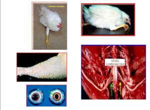
Marek’s disease is a lymphomatous disease of domestic chicken, caused by herpes virus. In the nervous form there are no gross lesions. However, lesions on nerves are only visible under microscope. In viscerous form, greyish white tumours are found in the ovaries, liver, spleen, kidney, heart and other organs. Lesions in the eye appear as loss of pigmentation of iris due to infiltrating lymphocytes with irregular pupil causing blindness called ‘grey eye’.
New castle disease
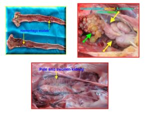
It is an acute contagious disease of domestic chickens caused by Type 1 Avian Paramyxovirus. Post mortem lesions include haemorrhagic ulcers on the mucosal lining of intestines, pinpoint haemorrhages on the tips of proventricular glands, haemorrhagic caecal tonsils, congestion and haemorrhages in trachea, air sacculitis, egg peritonitis among layers and marked congestion of pectoral muscles.
Fowl cholera
It is one of the most virulent and highly septicemis disease caused by Pasteurella multocida. Lesions seen in per-acute and acute cases include marked congestion of the carcass, multiple petechiation throughout viscera, multiple pin point necrotic foci in the liver. Presence of free yolk in body cavity, edema of lungs, pneumonia and perihepatitis are observed in layer birds. In chronic form we see caseous arthritis of hock joints, swelling and induration of one or both wattles, caseous exudates in the middle ear.
Pullorum disease
This disease mostly affects young chicks and is caused by Salmonella pullorum. Post mortem findings include unabsorbed yolk sac, peritonitis, congested lungs, dark, swollen and haemorrhagic liver and typhlitis. In growers, arthritis of hock joint is seen with the presence of lemon or orange coloured gelatinous material. In adults, abnormal ovary with ova, peritonitis, arthritis and pericarditis are seen.
Fowl typhoid
This is also known as Infectious Enteritis and is caused by Salmonella gallinarum. In acute phase, we see lesions like septicaemic and jaundiced appearance, congested skeletal muscles; swollen, friable and dark red liver with coppery bronze sheen surface, enlarged spleen, catarrhal enteritis, characteristic dark brown bone marrow. In chronic phase, emaciation, anaemia, focal necrosis in heart, intestine, pancreas and liver are seen. Greyish white necrotic foci seen in the myocardium, mucosa of intestine and pancreas, pericarditis with morbid yellow fluid in the pericardial sac, retained yolk which may subsequently rupture in layers.
Colibacillosis
It is caused by Escherichia coli. Post mortem findings include air sacculitis, perihepatitis, pericarditis, and peritonitis. Liver, spleen, lungs and kidneys are dark and congested; air sacs are thickened, opaque and white. Fibrinous pericarditis with thickened pericardial sac adhering to surface of the heart is a characteristic feature of this disease. The surface of the liver is also covered with gelatinous, greyish fibrinous material.
Aspergillosis
It is also known as Brooders Pneumonia or Pneumomycosis caused by Aspergillus fumigatus. In post mortem, presence of small, white caseous nodules measuring approximately 1 mm in diameter are seen scattered throughout the lung tissue and also air sacs. Lesions in brain tissue appear as white to yellow circumscribed areas.
Candidiasis
This is also known as Moniliasis or Thrush. It is caused by Candida albicans. Lesions are common in crop and to lesser extent or frequency in mouth, oesophagus and proventriculus. In acute cases, crop lining will be thickened and opaque. In chronic cases, the crop reveals raised white patches of ulceration and still in most severe case, there will be turkish towel appearance of crop with thickening. Mouth, oesophagus and proventriculus reveal ulceration and inflammation.
Coccidiosis
It is an expensive and very common disease of poultry that cause significant economic loss with universal importance and is caused by a protozoan parasite belonging to genus Eimeria. Lesions are mostly seen in intestinal tract in the form of haemorrhages and massive swellings. Serous surfaces reveal white focal lesions and haemorrhagic spots. Intestinal contents consist of blood and mucus mixed with necrotic material which is described as gruel like mass.
Main avian diseases found in India
MAREK’S DISEASE (MD)
Marek’s disease is a neoplastic disease caused by an oncogenic herpesvirus. The virus infects lymphocyte cells that, in some cases, will become tumour cells and infiltrated in different organs and tissues of the animal.
It has a worldwide distribution, so it could be said that all birds end up being exposed to the virus, although only a few cases develop the disease, usually the ones that are still very young and are not vaccinated or if there is a vaccinal failure.
Infected animals without symptoms are of primary importance since they are the ones that cause greater economic losses, due to poor growth of the animals, loss of uniformity and quality of the carcass, reduction in egg production, greater susceptibility to other diseases and worse response in other vaccinations.
Symptomatology depends on the location of the tumours and appears in the rearing animals that are almost ready for production. There are three types of presentation of the disease: cutaneous, nervous or visceral.
The cutaneous presentation appears as nodules at the level of the follicles of the feathers.
Nervous presentation shows different signs depending on the peripheral nerve in which the lymphocytic infiltration occurs, although the most frequent is to observe flaccid paralysis of the legs due to unilateral affection of the sciatic nerve or blindness due to the effect of infiltration on the nerve.
The visceral presentation with nodular lesions is what causes the general disorders and death of the animal.
NEWCASTLE DISEASE (RD)
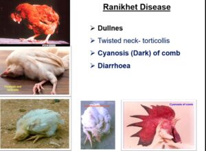
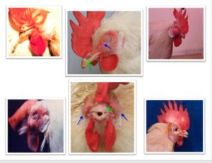
It is a highly contagious disease, caused by an avulavirus, in which the usual form is a respiratory condition, but other main clinical signs may include nervous manifestations (depression, drooping wings, twisted head or paralysis), diarrhea and swelling of the eyes and the neck.
Its importance lies in the mortality and the low productive yield an outbreak could cause, since it may lead to a partial or total stop in egg production and rough or thin egg shells.
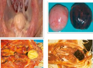
This pathology, also known as Ranikhet disease in India, shows diphtheritic hemorrhagic lesions of the alimentary tract, mainly in the ceca and cloaca, in its viscerotropic presentation. It is also common to find edema and petechiae in the gizzard and hemorrhagic ovaries.
INFECTIOUS LARYNGOTRACHEITIS
It is a respiratory disease caused by a herpesvirus that could appear in different presentations: peracute, subacute or chronic. Due to this, clinical signs vary from an extreme severity, with deaths by asphyxia, to a very mild symptomatology, that is not possible to differentiate from other respiratory diseases.
This virus causes fibrinous and hemorrhagic infiltration of the respiratory tract, so the presence of blood in the trachea, sinuses and oral cavity makes breathing difficult. In the postmortem examination, fibrinous, necrotic, caseous and/or diphtheritic plaques and plugs are found in the trachea, larynx and mouth.
GUMBORO DISEASE (IBD)
Gumboro disease, also known as infectious bursal disease (IBD), is an acute viral infection, caused by a birnavirusthat, as one of its names indicates, it mainly affects the Bursa of Fabricius, destroying the immature B lymphocytes of young animals.
Clinically, it is observed in chickens between 3 and 6 weeks old, which show depression and diarrhea. This diarrhea is the cause of dehydration, that affects the kidney and may cause the death of the animal.
Younger birds suffer from a subclinical disease causing immunosuppression, and thus increasing susceptibility to secondary infections and decreasing effectiveness of vaccines and productive parameters.
In cases showing clinical signs of the disease, diffuse hemorrhagic lesions are often observed in pectoral muscles and thighs. Hemorrhages and erosions may also appear at the level of the proventriculus-gizzard junction, as well as different degrees of nephritis or nephrosis.
Lesions at the level of the Bursa of Fabricius are variable and depend on the evolution of the disease. Initially, it is enlarged and edematous, around 5 days post-infection it returns to its normal size, although it can be hemorrhagic until it finally atrophies.
INFECTIOUS BRONCHITIS (IB)
This viral disease, exclusive of chickens, is confined to the respiratory system and the urogenital tract. There are different strains of the virus, with greater or lesser affinity for the systems named before. This virus has high mutagenic capacity, hence the main difficulty to fight against it, because vaccination does not guarantee to be resistant to the disease.
Most visible signs are those of the upper respiratory tract (sneezing and nasal discharge), but the condition of the ovaries is of greater importance, since it produces a marked decrease in the egg production and an increase in poor quality eggs (deformed or without shell). When kidneys are affected, a significantly increase in water consumption may appear, resulting in watery stools and wet litter.
According to all that it is mentioned above, the most frequent lesions caused by this disease are: serous, hemorrhagic, catarrhal or caseous exudate in trachea; pneumonia and/or opaque air sacs with possible caseous material; atrophied ovary and inflamed oviduct and interstitial nephritis, where kidneys are enlarged and pale.
INFECTIOUS CORYZA (IC)
Infectious coryza is a highly contagious bacterial disease produced by Avibacterium paragallinarum. It shows mainly rhinitis and infraorbital sinusitis, seen as facial edema in this area.
Conjunctivitis and abundant nasal and ocular secretion that may cause eyelids adherence could also appear. Usually, this state is accompanied by decreased consumption of food and water, and in laying birds, by variable reduction of egg production. Low respiratory tract infection rarely occurs.
CHRONIC RESPIRATORY DISEASE (CRD)
It is a contagious disease, with slow development and prolonged course, caused by bacteria of the genus Mycoplasma spp. Affected animals develop respiratory symptoms such as sneezing, nasal discharge or dyspnea.
In broilers, mortality rate may be low in cases without complications, but could reach 30% in cases complicated with other bacteria or viruses. There are also collateral losses due to stunted growth and sacrifice before reaching the term. Mortality can be low in layers; however, it causes a drop in the laying rate.
Lesions that appear are very variable, depending on whether there are other concomitant infections or not and on the mycoplasma agent itself. Sinusitis, tracheitis, air sacculitis, thickening and turbidity of the alveoli, exudative accumulations, fibrinopurulent pericarditis and perihepatitis could be observed.
FOWL CHORERA or AVIAN CHOLERA
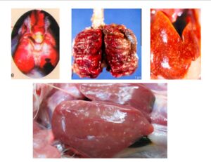
It is a contagious bacterial disease, caused by Pasteurella multocida, which affects adult birds or birds in the period of sexual development, that consists of 3 clinical presentations:
- PERACUTE FORM: Development of sudden deaths without previous symptoms. It is not frequent nowadays due to the immunization of breeders.
- ACUTE FORM: It has a course of 1-2 days, where the birds show anorexia, fever, depression, profuse white or grayish diarrhea, respiratory difficulty with abundant mucus, and cyanosis visible on crests and wattles.
- CHRONIC FORM: Animals remain sick and cachectic for a long time, with purulent or caseous inflammation of the wattle and possible subcutaneous abscesses.
Mortality and morbidity are variable and may affect a high percentage of animals. Acute cases that survive tend to evolve towards chronicity. Avian cholera causes very high economic losses due to high mortality, decreased posture and reduced fertility of hatching eggs. Outbreaks occur during low temperatures and high humidity (late summer, autumn and winter).
Necropsy findings seen in an acute outbreak lesions look like hemorrhagic septicemia, with petechiae and generalized hemorrhages in organs and skin, congestive spleen without splenomegaly, milliar necrosis in the liver and edematous lungs, sometimes with small purulent grayish areas. Caseous masses may appear in the air sacs and/or in the peritoneum in chronic presentations.
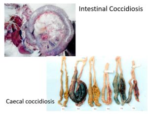
COCCIDIOSIS
Avian coccidiosis is a parasitic disease caused by protozoa of the genus Eimeria, originating a clinical or subclinical process characterized by bloody diarrhea and decreased production. There may also be a certain degree of dehydration, blood stained cloaca and anemia.
Destruction of the epithelial cells and the villi is what leads to a malabsorption syndrome that causes the loss of productivity of the infected animals. In addition, these lesions enable the action of other pathogens, such as Clostridium.
There are 7 species of Eimeria identified as pathogens of chickens, all located in the small intestine, except E. tenella, which is located in the caecum. Because of that, different lesions could be observed depending on the type of Eimeria and its virulence. Most of clinical outbreaks are due to mixed infections of several species of Eimeria and, therefore, lesions are observed in different areas of the intestine.
This protozoon causes enteritis of different severity, usually hemorrhagic, that is visible externally as petechiae. Species located in the cecum cause hemorrhagic typhlitis, with fresh or clotted blood mixed with fibrous exudate.
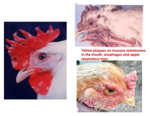
FOWL POX or AVIAN POX
It is a moderate to severe viral disease, with a slow development, caused by a poxvirus.
This virus induces a rapid growth of superficial layers in the skin and mucous membranes that form masses of proliferative tissue. The disease has two presentations depending on its location: cutaneous or “dry” from, on the skin without feathers, or diphtheric or “wet” form, in the alimentary and upper respiratory tract. Both could evolve alone or simultaneously. Diphtheria injuries are more serious, as they can cause death from suffocation or starvation.
Infection may occur at any time of the year, however, is believed to exist an association between outbreaks of the disease and climatic conditions favorable to the abundance of mosquitoes, with rains and warm temperatures.
Lesions vary according to the stage of development: papules, vesicles, pustules, or crusts, mainly in the region of the head. Diphtheric lesions are yellowish or whitish plaques that grow on the mucous shells of the nasal and buccal cavities, sinuses, larynx, pharynx, trachea or esophagus.
CONCLUSIONS
After describing each disease, it could be observed that their common fact is that they all lead to a decrease in decrease in productive parameters, either because broilers do not grow at the expected rate or because egg production is not appropriate for the week of the laying period and/or the quality of the egg is worsened.
The best way to solve these problems is to anticipate them. Applying the three essential points mentioned below will allow us to prevent the outbreak of a disease or will improve our control on it:
- Apply a management, including environmental and animal density monitoring, suitable vaccination plan, etc.
- Maintain adequate hygiene conditions and control of facilities that prevent the entry of wild animals.
- Establish an appropriate nutritional plan for each period and ensure that this feed is stored in the right conditions.
Population growth, urbanization and the rise of income has increased the demand for animal protein at an exponential rate. The commercial poultry population has increased by 4.5% over the previous census and backyard poultry has increased by 45.8%. An increase of 16.8% has been observed in the total poultry population of the country. Thus, we must provide quality treatment to the poultry farmers and stakeholders in order to popularize poultry farming in India. This can serve as a revolution to cater the protein needs of the citizens. Post mortem examination helps in proper diagnosis of disease and in control of death in case of outbreaks and further treatment and prevention.
POST-MORTEM LESIONS OF IMPORTANT ANIMAL & POULTRY DISEASES
Necropsy findings in major poultry diseases

Necropsy findings in major poultry diseases
POST-MORTEM LESIONS OF IMPORTANT ANIMAL & POULTRY DISEASES
Compiled & Shared by- Team, LITD (Livestock Institute of Training & Development)
Image-Courtesy-Google
Reference-On Request.