


Recent Advances in Fine Needle Aspiration Cytology (FNAC) for Diagnosis in Veterinary Sciences
Dipesh Kumar Dabi, Pranesh M and Geeta Devi Leishangthem
Department of Veterinary Pathology, College of Veterinary Science, Guru Angad Dev Veterinary and Animal Sciences University, Ludhiana, Punjab, India.
*Corresponding Author: dipesh692001@gmail.com
Abstract
Fine Needle Aspiration Cytology (FNAC) has long been an essential tool in veterinary diagnostics due to its simplicity, cost-effectiveness, and minimally invasive nature. However, recent scientific and technological innovations have transformed FNAC into a highly sophisticated diagnostic approach. This review highlights the recent advances in FNAC techniques in veterinary science, examining each development’s advantages and disadvantages. From digital cytology, artificial intelligence (AI), and machine learning applications to molecular diagnostics, imaging-guided FNAC, and the emergence of standardized protocols, this article presents a comprehensive overview of how FNAC is evolving into a more reliable, precise, and informative diagnostic modality.
Introduction
FNAC is a quick, minimally invasive procedure used to assess lumps or masses by aspirating cells through a fine needle, providing cytological diagnosis without the need for surgery. In veterinary medicine, accurate and rapid diagnosis of tumors and other lesions is crucial for effective treatment planning. It offers a minimally invasive method to collect cellular material for microscopic evaluation. Whether for diagnosing a suspicious lump or staging a tumor, FNAC helps to deliver rapid answers without surgical intervention. Despite its early success, FNAC traditionally faced challenges such as limited sampling accuracy and subjective interpretation. Recent advances aim to overcome these hurdles, enhancing diagnostic yield, reliability, and clinical utility. This article explores these innovations, emphasizing their practical implications in veterinary science.
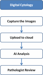
Digital Cytology and Artificial Intelligence
Digital cytology offers real-time analysis with high-resolution images of FNAC smears, enabling further tele-analysis and even pattern recognition through AI within seconds and can tell minutiae differences. Conventional FNAC vs. Digital Cytology as a novel approach, digital cytology and AI in FNAC have the potential to revolutionize the field of cytopathology through improved accessibility, efficiency, training, and diagnostic performance.
Advantages:
Enables remote consultations (telecytology)
Reduces human error
Accelerates diagnosis
Disadvantages:
Requires expensive equipment
Needs stable internet
Initial setup cost is high
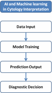
Machine Learning Algorithms in Cytology Interpretation
Machine learning (M L) models
Machine learning models are trained on thousands of annotated cytology images to recognize malignancy patterns and cellular features for accurate diagnosis.
Advantages:
Improves diagnostic accuracy
Reduces pathologist workload
Learns continuously from new data
Disadvantages:
Requires vast datasets
Cannot fully replace human judgment
Risk of bias if poorly trained
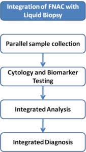
Integration of FNAC with Liquid Biopsy Techniques
Liquid biopsy analyzes biomarkers from blood or body fluids, complementing FNAC findings. It serves as a valuable complement to FNAC by providing molecular-level insights, especially for tumors that are difficult to access.
Advantages:
Provides molecular and cytological data
Useful for inaccessible tumors
Monitors tumor recurrence
Disadvantages:
Specialized lab required
Risk of false positives/negatives
Costly procedure
Molecular Diagnostics from FNAC Samples
Material from FNAC can undergo PCR and NGS for detecting genetic abnormalities.
Advantages:
Enables precise tumor typing
Supports personalized treatments
Detects minimal residual disease
Disadvantages:
High technical complexity
Costly
Not feasible everywhere

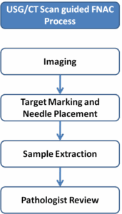
Imaging-Guided FNAC (Ultrasound and CT Assistance)
Ultrasound or CT imaging helps guide needle placement precisely during FNAC. This technique is becoming very popular in well-developed clinics as it allows accurate sampling of deep-seated or small lesions, minimizes complications, and improves diagnostic yield, especially in challenging veterinary cases.
Advantages:
Targets accurate lesion areas
Minimizes sampling errors
Reduces complications
Disadvantages:
Equipment expensive
Trained personnel needed
May not be available in rural setups
3D Cytology Imaging and Predictive AI Models
Emerging era of 3D cytology imaging and AI models can predict tumor behavior and progression in a very precise and non-invasive manner, significantly enhancing early diagnosis and treatment strategies in veterinary oncology
Advantages:
Better visualization of cells
Early tumor prediction
Highly individualized therapy
Disadvantages:
Still experimental
Needs heavy computational resources
Standardization of FNAC Protocols and Training Programs
Standardized protocols and training ensure consistency in FNAC practices for that a guidelines should be issued with detail description of all the important veterinary diseases and tumors so it will be easy for veterinarians to easily diagnose the anomaly.
Advantages:
Reduces observer variability
Improves diagnostic quality
Boosts training outcomes
Disadvantages:
Implementation requires time
Resistance from traditionally trained practitioners
Table 1. Comparison of FNAC Technological Advancements
| Innovation | Advantages | Disadvantages |
| Digital Cytology | Remote access, fast diagnosis | Expensive equipment, internet-dependent |
| Machine Learning | High accuracy, reduces workload | Needs large datasets, bias risk |
| Liquid Biopsy | Molecular + cytology data | Costly, risk of errors |
| Molecular Diagnostics | Precise typing, personalized therapy | Technically complex |
| Imaging-Guided FNAC | Precise targeting, fewer errors | Needs expensive tools |
| 3D Imaging + AI | Early prediction, better views | Still experimental |
| Standardized Protocols | Consistency, better training | Slow adoption |
Conclusions:
Recent advances in Fine Needle Aspiration Cytology have significantly expanded its diagnostic potential in veterinary science. Innovations like digital cytology, AI, machine learning, liquid biopsy integration, molecular diagnostics, and imaging guidance have transformed FNAC from a basic tool into a sophisticated, multi-parametric diagnostic modality. Each of the advancement brings clear advantages but also poses challenges that require careful adaptation and continuous training. By integrating these advancements, veterinarians can ensure earlier, more accurate diagnoses and optimized therapeutic interventions, improving the overall health outcomes for animal patients.
References
Smith, J., Brown, R. and Patel, K. (2021). AI in Veterinary cytology: Revolutionizing tumor diagnostics. Veterinary Pathology Journal. 58(4): 499-508.
Jones, M., Taylor, P. and Stevens, L. (2022). Machine learning in Veterinary Oncology: Opportunities and challenges. Journal of Veterinary Diagnostic Investigation. 34(2): 225-234.
Davis, R., Hamilton, A. and Green, S. (2023). Liquid biopsy applications in Veterinary cancer diagnosis and monitoring. Veterinary Oncology Reports. 12(1): 15-24.
Thompson, A., Wilson, D. and Carter, B. (2020). Molecular diagnostics in veterinary oncology: Current perspectives. Journal of Small Animal Practice. 61(5): 263-271.
Wang, Y., Lin, T. and Zhao, H. (2021). Role of imaging modalities in guiding fine-needle aspiration in veterinary tumors. Veterinary Radiology & Ultrasound. 62(3): 251-259.
Adams, K., Spencer, M. and Lewis, J. (2023). Standardization in veterinary cytopathology: Challenges and solutions. Veterinary Clinical Pathology. 52(1): 6-14.