

SUBMANDIBULAR AND SUBLINGUAL SALIVARY MUCOCELES IN DOGS
Dr. N. Krishnaveni*, M. Kabilan and D. Manikandan
Department of Veterinary Surgery and Radiology
Veterinary college and research Institute, Tirunelveli
Tamilnadu Veterinary and Animal Sciences University
*corresponding author e-mail ID: veninarayanan110@gmail.com
Assistant Professor,
Department of Veterinary Surgery and Radiology
Veterinary college and research Institute, Tirunelveli
Introduction
Salivary gland disorders have rarely been described in dogs and cats, with only a 0.3% overall prevalence. The mucocele is a non-epithelial, non-secretory linning, mainly consisting of connective tissues and capillaries (Karbe and Nielsen, 1996). A Salivary mucocele (sialocele), salivary cyst, honey cyst is a collection of saliva that has leaked from damaged salivary gland or duct surrounded by granulation tissue. A sublingual mucocele is a collection of saliva in the sublingual tissue caudal to the openings of the sublingual and mandibular ducts. Salivary mucocele are not cyst. Dogs are more frequently affected than cats (Dunning, 2003). All breeds are susceptible but some reports indicate that poodles, German shepherds, dachshunds, Australian silky terriers are more commonly affected. An animal of any age may develop a mucocele but males are more prone.
The interior of the mucocele suppurates and gradually closes by granulation. The cause of salivary mucocele is rarely identified, although blunt trauma, foreign bodies sialolith have been suggested. Tearing of the salivary gland or duct is resulting in leakage of saliva into the surrounding tissues. Saliva irritates the tissue and causes inflammation. The swelling may be firm painful initially but the animal is usually asymptomatic. Granulation tissue forms in response to inflammation and prevents the saliva from migrating further. The diagnosis of salivary mucocele is based primarily on the history, location, clinical signs, cytology findings, radiographs and histopathologic examinations.
Etiology
The causes of salivary mucocele are poorly understood, although blunt trauma, foreign bodies, sialoliths and trauma during chewing have been proposed. It arises most commonly from the sublingual salivary gland, either from individual units of the polystomatic portion or from the duct of the monostomatic portion in dogs (Brown, 2007). Other suggested causes are haematomas, neoplasia and recent oral surgery (Olimpo et al., 2023).
Clinical findings
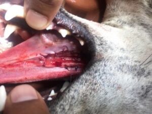
Clinical findings depend on the location of the mucocele. Asymptomatic animals usually present with the history of a gradually developing, fluctuant, painless mass. The patients with sublingual mucocele (Fig.1) may have abnormal prehension and oral bleeding, which is caused by trauma during chewing. Swelling in the oro-pharngeal area may cause abnormal tongue movements and interfere with eating (dysphagia) or breathing (respiratory distress).
On physical examination parotid and mandibular glands are easily palpated without discomfort. Sublingual gland is palpable in sedative patients. Most mucocele are soft and fluctuant whereas tumours and abscess are usually firm. Mucocele are painless except during the acute phase of swelling. When the mucocele are located on the ventral midline or inter mandibular space (Fig.2), examining these animals in dorsal recumbency to gravitate the affected side. In traumatise mucocele blood tinged saliva may be seen. Periorbital facial swelling, enophthalmos and periocular pain are the signs of zygomatic mucocele.
Diagnostic approach
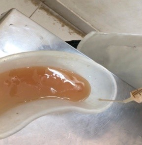
Based on history, physical examination and result of exploratory fine needle aspiration the case can be diagnosed as salivary mucocele. The diagnosis of salivary mucocele is usually confirmed based on aspiration of the identified mass Torad and Hassan, 2013). Paracentesis should be performed under aseptic conditions to prevent infections of the mucocele. Aspiration of a clear yellowish or blood tinged , ropy, mucoid fluid (Fig.3) with a low cell count is consistent with saliva. Staining a smear with periodic acid Schiff (PAS) confirms the presence of saliva.
Retrograde sialography (i.e., injecting iodinated, water soluble contrast agent into a salivary duct) can be used to further confirm diagnosis and identify the affected gland for surgical planning. Advanced imaging modalities such as ultrasonography, MRI, and CT can also be used to provide additional details about soft-tissue characteristics and the extent of involvement. These could be helpful for differentiating this disease from other causes of submandibular swelling in dogs (Yasonu et al., 2011).
Differential diagnosis
Sialoadenitis, sialadenosis, salivary neoplasia, sialolith (calcium phosphate or carbonate), cervical abscess, foreign body, hematoma, cystic or neoplastic lymph nodes, tonsil cyst, thyroglossal cyst, cystic Rathke’s pouch, or branchial cysts may cause swellings in the same region as mucoceles. Histopathologic examination is necessary to diagnose salivary gland tumors and to differentiate a congenital cyst from a mucocele. Congenital cysts have an epithelial lining, whereas mucoceles are lined by granulation tissue.
Therapeutic strategy
Emergency aspiration may be necessary for the animal in respiratory distress. Since it has abscessation or fibrosis are the complication, repeated drainage or injection of cauterizing or anti inflammatory agents are contraindicated. Complete excision of the involved gland or duct complex and the drainage of the mucocele are curative.
Surgical Technique
- Marsupialisation is the process of incising a mucocele and suturing the edges to the mucosa (Fig.4)
- Excision of the affected entire salivary gland complex.
Postoperative care and assessment
Change bandages daily if a Penrose drain has been placed. Depending on the amount of drainage, remove the drain 24 to 72 hours after surgery or when drainage is minimal. Allow the drain site to heal by second intention. Soft food should be fed for 3 to 5 days after ranula marsupialization or drainage of pharyngeal mucoceles.
Complications
Seroma formation, infection, and mucocele recurrence. Seromas can form in the dead space created by removal of the glands; they typically reabsorb and do not need aspiration or drainage. Mucoceles recur if the side of mucocele origin was misdiagnosed or if inadequate gland was not excised. Attention to anatomic detail during surgery will minimize recurrence and complications associated with salivary gland excision.
CONCLUSION
In rare cases a mucocele resolves without surgery. The prognosis is excellent if the disease is accurately diagnosed and excision is complete.
Reference
Torad, F.A and E.A. Hassan. 2013. Clinical and ultrasonographic characteristics of salivary mucoceles in 13 dogs. Vet Radiol Ultrasound, 54 (3): 293-298.
Yasonu H, H .Nagai, Y .Ishimura, T .Watanabe, H .Yamasaki, H .Anayama, Y. Takai, H. Yamauchi, Y. Hara, F. Murai and H. Kandori. 2011. Salivary mucocele in a laboratory beagle. J. Toxicol. Pathol. 24 (2): 131–135.
Olimpo, M.,E. Ilaria Ferraris, L. Parisi, P. Buracco, S. G. Rizzo, D. Giacobino, A. Degiovanni, L. Maniscalco and E. Morello. 2023. Diagnostic Findings and Surgical Management of Three Dogs Affected by Osseous Metaplasia Secondary to a Salivary Mucocele. Animals (Basels) 13 (9): 1550.
Dunning, D. 2003. Oral Cavity. In Textbook of Small Animal Surgery; Slatter, D.H., Ed.; Saunders: Wynnewood, PA, USA, pp.553–572.
Brown, C.C, D.C. Baker and I.K. Barker. Alimentary system. 2007. In: Pathology of Domestic Animals, 5th ed, Volume 2. M.Grant Maxie (ed.) Elsevier Limited, Philadelphia pp.32–33.
Karbe, E. and S.W. Nielsen. Canine ranulas, salivary mucoceles and branchial cysts. 1966. J Small Anim Pract. 7: pp 625–630.
 |
 |
||
| Fig.1 Sublingual mucocele (Black arrow) in a dog | Fig2. Submandibular sialocele (Red arrow) in a dog | Fig. 3 On aspiration, clear yellowish or blood tinged , ropy, mucoid fluid | Fig.4 Marsupialisation technique for ranula |