


SUCCESSFUL MANAGEMENT OF MYIASIS WOUND IN TAIL OF A DOG
Ankit Kumar1 Bhavna Dabas2
1INTERNEE STUDENT V.C.R.I. NAMAKKAL, TANUVAS
2THIRD YEAR STUDENT V.C.R.I. NAMAKKAL, TANUVAS
Email- ankitaiims357@gmail.com
Mobile no.- 8292265530
Abstract
Myiasis is the infestation of live human and other vertebrate animals with dipterous larvae which at least for certain period feed on the host’s dead or living tissue, liquid body substances or ingested food. It mostly occurs during the fly season. The present case report is of a 1-year-old dog infested with maggots in coccygeal region. The dog was successfully treated with rinsing by normal saline solution, turpentine oil, antiseptic solution and ivermectin injection.
Keywords: dog, maggot, wound, healing, therapeutic management.
Introduction
Myiasis or maggot wound refers to the parasitic infestation of tissue of a living vertebrate animal by larvae of the order Diptera (true flies) that grow inside the host while feeding on its dead or living tissue causing more or less traumatizing injury depending on the relationship of the larvae with the host and the location (Hall and Farkas, 2000; Francesconi and Lupi, 2005; Robbins and Khachemoune, 2010). Many different species of flies can be associated with this type of myiasis, the most common belonging to the family Calliphoridae (blow flies), followed to a lesser extent by the Sarcophagidae (flesh flies), Muscidae (house flies) and Phoridae (scuttle flies) (Day et al., 2000, Huntington et al., 2008 and Ibrahim et al., 2008). Clinically, myiasis can be classified based on the area of the body affected by the maggot or subsequent development in the host (Francesconi and Lupi, 2006). Therapeutic management of the myiasis wounds includes mechanical removal of maggots or by application of wound cleansing agents and/or a combination of both followed by general wound management. Cleansing agents are used to irrigate myiasis wounds in an attempt to kill the larvae, and/or to flush out any living or dead larvae from the wound, thus reducing the likelihood of secondary infection. Reported cleansing agents used in the treatment of cutaneous myiasis include saline solution, hydrogen peroxide (3%), diluted chloroform (with 5–15% light vegetable oil or milk) (Verettas et al., 2008, Caissie, 2008 and Szakacs et al., 2007), iodine, Dakin’s solution, ethyl chloride (Bedford et al., 1933], Chlorhexidine, ivermectin in a propylene glycol solution and water (Huntington et al., 2008 and Victoria et al., 1999). This case reports a case myiasis wound due to traumatic injury in tail of a non-descriptive dog.
Case history and Clinical observations
A 1-year-old male dog was presented at Government Veterinary Dispensary, Ernapuram Namakkal, Tamil Nadu with history of traumatic injury in the tail region 20 days back. Clinical examination of the animal revealed presence of maggots at the tail with slight bleeding. All the vital parameters were in the normal range. Clinical findings confirmed that dog was infested with myiasis wound in coccygeal region.
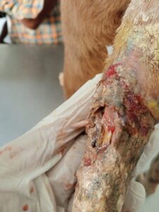
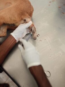
Dog infested with larvae in tail region. Flushing with Normal Saline using 10cc syringe.
Therapeutic management and Recovery
The hairs around the maggot wound were clipped. The wound was then rinsed with normal saline to wash away the dead maggots present on the surface of wound. Rinsing was done under pressure using 10cc syringe and repeated 3-4 times. The remaining maggots were gently wiped away or removed with a pair of tweezers or artery forceps. Gauze bandage soaked with turpentine oil was kept on the open wound for about 20 minutes to draw out and kill maggots that have burrowed deeply into the wound. The wound was again rinsed with normal saline water to wash away the remaining maggots. Ivermectin injection @0.3 mg/kg body weight was administered by subcutaneous route. Supportive therapy of Enrofloxacin @10mg/kg body weight, and Tramadol @2mg/kg body weight as analgesic was administered. Aluspray-AWD was advised topically. After 1 week of treatment, the dog recovered fully indicated by absence of maggots and bleeding in coccygeal region.
For animal with traumatic wound as reported in this case, proper treatment of the wound needs to be done promptly to prevent further complications. Clinical presentation of myiasis may vary depending on the specific cause and the body part affected. In the present case, the dog was presented with traumatic injury in the tail region where maggots were seen on the wound. Application of a toxic substance to the larva and egg, production of localized hypoxia to force the emergence of the larva, mechanical or surgical removal of the maggots from the site of infestation and surgical debridement are the various therapeutic measures indicated for the management of various types of myiasis (Francesconi and Lupi, 2006). After removal of the larvae and dead tissues, the wound was dressed daily with antiseptic solution, administration of Ivermectin and systemic administration of antibiotic against secondary bacterial infection were done in this case and in agreement with Falish (2012) and Abdullah et al., (2015). The case presented with maggot wound in the dog was clinically managed effectively by standard way of the treatment of myiasis and there was uneventful recovery.
References
Bennet, R. (1988). Fundamentals of cutaneous surgery. St. Louis: C.V.Mosby:Pp.778.
Bowler, P., Duerden, B. and Armstrong, D. (2001). Wound microbiology and associated approaches to wound management. Clin.Microbio.Rev. 14: 244-269.
Caissie, R. (2008). Cutaneous myiasis: diagnosis, treatment and prevention. J. Oral.Maxillofac. Surg. 66: 560–568.
Casu, R.E., Pearson, R.D., Jarmey, J.M., Cadogan, L.C., Riding G.A. and Tellam, R.L., (1994). Extretory/secretory chymot rypsin from Lucilia cuprina: purification enzymatic specificity and amino acid sequence deduced from mRNA. Insect Molecular Biology. 3: 201–211.
Hall, M.J. and Farkas, R. (2000). Traumatic myiasis of humans and animals. In: Contributions to a Manual of Palaearctic Diptera. Science Herald, Budapest. pp: 751-768.
Huntington, T.E., Voight, D.W. and Higley, L.G. (2008). Not the usual suspects: human wound myiasis by Phorids. J. Med. Entomol. 45: 157–159.
Victoria, J., Trujillo, R. and Barreto, M.(1999). Myiasis: a successful treatment with topical ivermectin. Int. J. Dermatol. 38: 142–144.
Zumpt F. Myiasis in man and animals in the old world. Butterworths, London. 1965; 267.