


SURGICAL MANAGEMENT OF ATRESIA ANI IN NEW BORN KID
Ankit Kumar1 Dharya Yadav2
1INTERNEE STUDENT V.C.R.I. NAMAKKAL, TANUVAS
2THIRD YEAR STUDENT V.C.R.I. NAMAKKAL, TANUVAS
Email- ankitaiims357@gmail.com
Mobile no.- 8292265530
Abstract
A five-day old kid was presented at the Veterinary Dispensary Eranapuram, Namakkal, Tamil Nadu with complaints of distended abdomen and inability to defecate. Type 1 atresia ani was diagnosed on physical examination. Surgical reconstruction is the only course of action for congenital atresia ani (kid) in new born animals
Key words: Atresia ani, congenital defect and non-descriptive kid.
Introduction
Atresia ani (imperforate anus) is a congenital abnormality characterised by the persistence of anal membrane resulting in a thin membrane covering the normal anal canal. It may be caused by genetic or environmental factors, or a combination of both; in many cases, the causes are unknown. The most common caprine environmental teratogens include toxic plants consumed by the dam and maternal-foetal viral infections during gestation. Congenital malformation sometimes leads to perinatal mortality, and it may also decrease maternal productivity and reduce the value of the defective neonates. Severe defects result in abortion of the kid. There are four major types of intestinal atresia. This report communicates a case of atresia ani in kid, which was successfully treated by surgical intervention.
Case history and Clinical observations
A five-day old kid, non-descript breed was presented at Veterinary Dispensary, Eranapuram, District Namakkal, Tamil Nadu (India) with the history of non – passage of faeces since birth. After birth, kid was normal to stand and suckling behaviour and later became dull and depressed. On clinical examination, signs of dull and depressed, anorexia, attempt of defecation and mild abdominal distention observed. Tenesmus and evidence of abdominal pain were manifested by restlessness. The case was diagnosed as atresia ani and handover for surgical intervention.
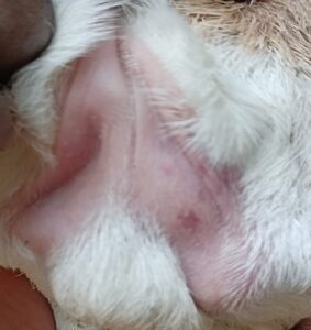
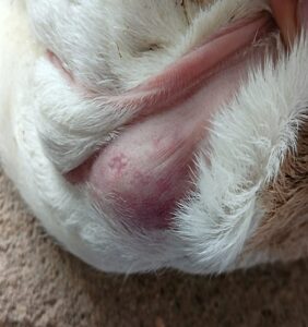
a)Absence of rectal opening with an b)Demarcation of anal area on caudal
intended open scar . compression of abdomen.
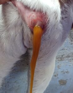
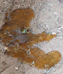
c)Photograph showing meconium after d)Photograph of meconium
incision into the rectum.
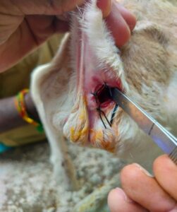
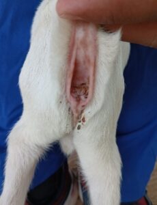
e)Photograph of constructed anal opening f)Photograph showing reconstructed
and placed sutures. anus after 2 months of recovery.
Treatment and Discussion
The kid was restrained properly, placed on sternal recumbency. The perineal area below the base of tail was prepared for routine aseptic surgery using chlorhexidine-gluconate antiseptic. Local infiltration anaesthesia was performed by using injection 2% lignocaine hydrochloride solution at the proposed site of incision. In present case, abdomen was compressed caudally for the anal area to be demarcated. Then a circular full thickness incision was made upon the bulged anus and the circular piece of incised skin was removed. The rectum was exposed after dissection of the perineal muscles therein and mild bleeding was occurring. The blind end of the rectum was brought to the level of anal sphincter (at incised skin) and fixed to the perineum after duly snipping the tip of the blind end of rectum meant for evacuation of the contents (meconium). This was done by putting four stitches (12 o clock, 3 o clock, 6 o clock and 9 o clock). The circumference of the rectal opening was sutured by application of interrupted sutures by using black braided silk # 2 between rectal mucosa and skin to make a permanent anal orifice and kid was stand normally with minimum tenesmus immediate after surgery. Post operatively, Ceftriaxone @5 mg/kg body weight for 3 days and Meloxicam @ 1 mg/kg body weight for 3 days were administered intramuscularly, followed by routine dressing with Povidone iodine and application of fly repellent ointment at the operative site till recovery. The sutures were taken off on 10th day post-operatively. Congenital defects and abnormalities presented in this study were recorded as sporadic cases and it may be due to genetic or environmental forces, or a combination of both, during the process of embryogenesis. Affected kids initially will stand and suckle normally after birth. The time of onset of clinical signs of disease may vary from 1 to 3 days. On collection of history of the owner did not see the kid passing the meconium or faeces was the main observation. The principal clinical signs of present case were straining, colic, tenesmus, depression, anorexia with moderate abdominal distention and not passing meconium. Atresia ani simply can be diagnosed by visual inspection of anal opening at perineal region or by limited digital palpation if a vestigial anal opening is present. The kid showed marked improvement in defecation with minimum tenesmus and active in nature within 2nd day of surgery and uneventful recovery occur within 10th post operative day. Four major types (type – I, II, IIIa & III b and IV) of intestinal atresia have been described by Raha et al…2007. Atresia ani is a type I atresia in which the mucosal blockage within the intestinal lumen. The present case of atresia ani of intestinal atresia is the simple form of agenesis without involving the other parts and similar findings in kid calves were reported. It was also found with other congenital defects reported by various authors like anus vaginalis, atresia ani et recti, atresia ani with vaginal-urinary bladder agenesis, atresia ani with scrotal anomaly and congenital recto-vaginal fistula with atresia ani. But in our study we observed atresia-ani alone. If the rectum ends blindly as a cul- de sac a short distance cranial to the anal membrane, the condition is called rectal atresia.
Conclusion
The diagnosis of atresia ani can be carried out on the basis of history, age and clinical examination of the animal. Prompt surgical intervention is required to relieve the animal from abdominal discomfort and for better prognosis.
Reference
[1]. Suthar D.N., S.R. Chaudhary, P.B. Patel, J.N. Mistry, J.B. Patel and S.S. Nerurkar (2010). Surgical management of atresia ani in a cow calf. Vet World, 3: 380-381.
[2]. Noden, D.M and Lahunta A (1985). The embryology of domestic animals, developmental mechanisms and malformations, Williams & Wilkins, London; 306-315.
[3]. Kilic N, Sarierler M. Congenital Intestinal Atresia in Calves (2004; 61 Cases (1999–2003). Revue de Medicine Veterinaire. 155(7):381-384.
[4]. Oehme, F.W and Prier, J.E (1974). Textbook of large animal surgery. Williams & Wilkins, Baltimore, U.S.A, 447-448.
[5]. Singh, A.P. (1989). Congenital malformation in ruminants- a review of 123 cases. Indian Veterinary journal 66:981-985.
[6]. Mallesh P, Sampath K, Raju G (2017) Successful management of atresia ANI (congenital defect) in non-descript calf: A case report. The Pharma Innovation 6(7): 337-338.
[7]. Leipold, H.W, Dennis S.M, Huston K (1971). Congenital defects of cattle: Nature, cause, and effect. Advances in Veterinary Science and Comparative Medicine 16:103-150.
[8]. Hossain, M.B. Hashim M. A. Hossain M.A. Sabrin, M.S. (2014). Prevalence of atresia ani in newborn calve and their surgical management. Bangl. J. Vet. Med.12 (1): 41-45.
[9]. Remi-Ademun B.D., Fale, M.S. Usman, B., Lawal, M. (2007). Retrospective study of atresia ani cases presented at Ahmadu Bello university Zaria, Nigeria.NVJ.28(1):48-56.