


THERAPEUTIC MANAGEMENT OF POST-PARTURIENT HEMOGLOBINURIA IN A BUFFALO
Shalini, G. phD pursuing
Department of Animal Reproduction, Gynaecology and Obstetrics (ARGO)
College of Veterinary and Animal Sciences (CVAS), Mannuthy -680651
Introduction
Eutocia refers to normal childbirth, while dystocia indicates challenging delivery. If dystocia produces short- or long-term consequences, the effort may become unprofitable for dairy producers, posing significant financial risks. It is one of the most dangerous obstetrical illnesses and requires immediate attention from obstetricians due to the large financial losses dairy producers incur. Dystocia may be influenced by a variety of characteristics, such as breed, the dam’s parity, the calf’s sex, birth weight, the size of the dam’s pelvis, the length of the gestation period, diet, year and calving season. Fetal dystocia is more common than maternal dystocia in terms of incidence and the causes of dystocia are usually classified as either fetal or maternal. It may lead to increased rates of periparturient illnesses such metritis, retained placenta, and uterine infections. Due to the significant time, expenses and resources required to ensure that a cow conceives through artificial insemination (AI) or natural means, dystocia can have a devastating financial effect on farmers. This is because it can result in calf morbidity and mortality, higher veterinary expenses, decreased productivity, decreased fertility and in the worst cases, damage or death of the dam.
Case Discussion
The Puducherry region has a greater prevalence of dystocia during the COVID-19 era. My top focus is managing the situations at night and accurately diagnosing and treating them to save the mother and fetus without any issues. Good practical competencies, sensitivity to the welfare of both the dam and the offspring, and a thorough grasp of normal parturition are necessary for the diagnosis and treatment of dystocia. About ten o’clock at night, a five-year-old cross-bred Jersey cow with a history of sporadic straining and a ruptured water bag was attempted to deliver the fetus, but the animal was unable to do so. At approximately 11 p.m., the owner called to clarify the circumstances. Then, in less than 15 minutes, I reached with my obstetrical instruments were brought to that location. The animal was observed to be lateral recumbent and straining during an extremely difficult parturition. To stabilize the animal, an epidural injection of lignocaine and the intravenous fluids was given. Fetus was found to be in breech presentation upon rectal examination and the tail was palpable. The fetus was live with the presence of rectal reflux. The breech presentation was rectified in accordance with obstetrical procedure and the live female calf was extracted by forced traction after a snare was placed on both hind legs (Fig. 1). The fetal membrane was expelled after two hours of corrected dystocia (Fig. 2). The recovery of the dam and fetus went smoothly.
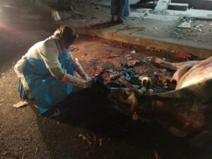
Fig 1: Dystocia caused by breech presentation corrected and a live female calf was removed
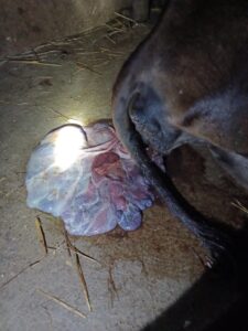
Fig 2: Placenta was expelled after two hours of fetus removed
A further episode of dystocia was reported on next evening. The six-year-old HF breed cow had a history of significant straining with the presence of one limb in the birth canal for the previous forty-five minutes without making any more progress. A rectal examination revealed a significant left lateral deviation of the head and neck together with a unilateral flexion of the shoulder. Snare was placed to one forelimb which was presented in the birth canal while under epidural anesthesia with lignocaine injection, then another forelimb was corrected. Snare was applied and both forelimbs were retropulsion to make space in the birth canal. Then, using a snare and an obstetrical blunt hook on the left inner canthus of the eye, the head was adjusted with light traction and the live female calf was removed by traction (Fig. 3).
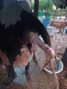
Fig 3: Dystocia caused by Postural defects of fetus
Conclusion
Abnormal fetal positions commonly manifest as posterior malpresentation, foreleg malposture, breech malpresentation or cranial malposture. The primary cause of dystocia is more frequently fetal than maternal cause. To preserve the health of the dam and fetus without any complications and to shield the farmers from financial loss, it is imperative that dystocia cases be diagnosed and treated immediately. Handled by farmer or attender should be strictly avoided because it leads to more complication to the dam and fetus. Introducing Jersey genes through crossbreeding in Holstein herds has the potential to reduce dystocia and enhance calf survival and health.