


TUBE CYSTOSTOMY AND URETHROTOMY IN BULLS
Dr Akash, PhD Scholar, Division of Surgery, ICAR-IVRI, Izatnagar, Bareilly, UP-243122

Email- anant.akash9@gmail.com
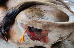
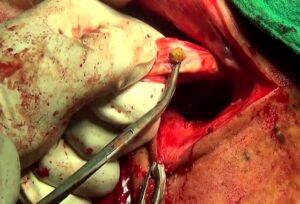
Tube cystostomy is a technique of urinary diversion in which a Foley’s catheter is placed into the urinary bladder lumen. The technique is used in the treatment of urethral obstruction, urethral rupture or ruptured urinary bladder. For tube cystostomy in bulls, a preoperative rectal examination for assessment of urinary bladder is done. In case of intact urinary bladder, tube cystostomy through blind approach can be performed in standing animal under sacrococcygeal epidural analgesia using 8-10 ml of 2% lignocaine. Left ischiorectal fossa is prepared for aseptic surgery. 3-4 ml of 2% lidocaine is infiltrated subcutaneously and into deep muscles before giving a nick incision at the site. 1-2 cm incision is given at the skin of left ischiorectal fossa for the insertion of catheter with the help of stylet. Before inserting the catheter, right hand is passed into rectum to ensure the path of stylet into urinary bladder and to avoid accidental puncture of rectum by stylet. A 16 FG foley’s catheter along with stylet is advanced towards the inflated bladder. After the assessment of the tip of catheter near bladder by right hand in the rectum, stylet is blindly pushed into the bladder. As the catheter enters into the urinary bladder, urine starts coming through catheter. Then the balloon of the catheter is immediately inflated with 30 ml of sterile water. The stylet is then retracted slowly and the external end of the catheter is then pulled gently. Urethrotomy should be performed after tube cystostomy in bullock to remove urethral caliculi. The animal is restrained in right lateral recumbency and post scrotal area is shaved and prepared for aseptic surgery. 2% lignocaine is infiltrated subcutaneously and deep intramuscularly 6-8 inches on midline and 2-3 inch behind scrotum. A midline incision of 3-4 inches is made behind the scrotum. Urethra is palpated on the ventral aspect and pulled out. Urethra is searched for the caliculi and after locating the caliculi a linear incision is given at urethra over the caliculi. Then caliculi is removed with the help of forceps. Urethra is catheterized with an elastic catheter. Urethral incision is closed with PGA No.2 suture and the catheter is anchored with the last suture. After reposition of urethra muscle layer is sutured with PGA No.1 suture. Skin incision is closed with polyamide No.2. The part of catheter outside the abdomen fixed with the skin with 3-4 sutures. Suture line is dressed daily with povidone iodine until healing. Post- operative antibiotic and analgesics should be given. Urolithiasis is a life threatening condition and need to be diagnosed and treated as early as possible with tube cystostomy followed by urethrotomy in bulls.
|
|
|