


A Mini Review on Diagnosis and Treatment of Lumpy Skin Disease (LSD) Menace in Cattle in India
Dr. Harshit Saxena¹
Kartikey Verma²
¹UG Scholar BVSc & AH, Intern, CoVSc & AH, DUVASU, Mathura, UP
²UG Scholar BVSc & AH, Third Year, COVAS, SVPUAT, Meerut, UP
________________________________________________________________
Abstract:
Lumpy skin disease (LSD) is an emerging infectious disease in Cattle in India caused by a virus of the family Poxviridae and genus Capripoxvirus. It is a vector borne disease transmitted by biting insects (For example, mosquitoes and biting flies). It affects the hide quality, milk production and can even cause death of the animal. Severe infections of the same can cause significant economic damage to dairy and beef cattle breeding. Vaccination can be done for the prevention of the disease, but studies have shown that it can cause various complications in animals in the form of liver dysfunctions and more severe cases may lead to embryonic mortality and abortions. Hence, the diagnosis and treatment of the disease is of utmost importance. This mini-review is specifically aimed at providing the latest diagnostic techniques and treatment practices to reduce further incidences in the future. It will also address the propagation and economic significance of the disease.
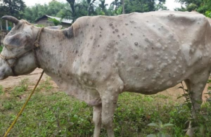
A Picture showing skin lesions of LSD in Cattle
(Courtesy: newindianexpress)
http://www.fao.org/3/u4900t/u4900t0d.htm
Keywords- Lumpy Skin Disease, Cattle, Livestock, India, Capripox virus, Ethnoveterinary Medicine, Veterinary Homeopathy.
Abbreviations- LSD– Lumpy Skin Disease
Introduction:
Lumpy Skin Disease Virus, which is also known as Neethling Virus was first isolated in South Africa [7]. This disease severely damages the skin of the cattle. The animal affect with this disease suffers from fever, enlargement of superficial lymph nodes, and the skin develops numerous nodules ranging from 2-5cm in size. Edematous swelling, if developed in the foot or limb, can cause lameness in the affected animal [8].
The genus Capripoxvirus is in the subfamily Chordopoxvirinae, family Poxiviridae. It contains 3 closely related viruses, namely Sheep pox (of sheep) and lumpy skin disease (of cattle) in addition to Goat pox virus (GTPV) which causes goat pox disease [11].
Epidemiology:
The country in which the disease was first reported was Zambia in the year 1949[15]. As of now, it has spread in almost all the countries in the African continent. Legal and illegal import of the live cattle is believed to be the major reason for the spread of LSD in Egypt and middle eastern countries [10].
In India, the first case of LSD was first reported in 5 districts of Odisha in the August of 2019. In total, 182 of 2,539 cattle were found to be affected with a morbidity rate of 7.1% and no casualty. Since then, it has spread all over the country including the states of Kerala, Uttar Pradesh, Madhya Pradesh and part of Chottanagpur region [9].
Lumpy skin Disease: A Menace:
The sudden appearance of skin lesions damages the skin, permanently affecting the meat industry of the country and its export. The skin lumps/ nodules start appearing within 2 days [2]. These nodules become harder with time. If necrosis starts in these areas, it can cause severe discomfort and lameness [6].
After 2-3 weeks, the necrosed area leaves a hole of full skin thickness which attracts secondary infections [2].
The occurrence of LSD also causes decrease in milk production and semen production. Permanent or temporary infertility may be caused in the affected bull [6].
LSD also causes liver and renal damage in the cattle [5].
The cattle suffer from anorexia, it becomes emaciated and severe cases may demand euthanizing the animals [13].
Diagnosis:
Etiology:
Lumpy skin disease is caused by an epitheliotrophic virus lumpy skin disease virus (LSDV) having a double-stranded DNA genome which belongs to the genus Capripoxvirus of family Poxviridae. This virus is shows similarities to the other Capripoxvirus species such as Sheeppox virus and Goatpox virus [1].
Host:
Lumpy skin disease has a narrow and specific host range which includes cattles and buffaloes and impala, giraffe, Thomsons gazelle, Arabian oryx and other animals [13].
Pathogenesis:
Lumpy skin disease virus enters the body of the hosts through skin. It can also enter through gastro intestinal tract. Virus then reaches the lymph nodes causing lymphadenitis. Virus causes skin lesions due to its rapid replication in specific cell and blood vessels walls with development of inflammatory nodules on skin. Skin nodules can turn to grey-pink with caseous necrotic cores. Circumscribed necrotic lesions may get ulcerated, it is referred as ‘sit-fasts [14].
Lesions:
Lesions begin as an area of reddening, progressing over two weeks to a papule, vesicle, pustule with exudation and scab formation. Healing is slow. A nodular form of the disease can also occur this resembles lumpy skin disease in cattle [13].
Post-mortem findings:
The epidermal and mucosal lesions described above will be apparent at post-mortem. Ulcerations may be found in the lining of the trachea and gastro-intestinal tract. Lung lesions consisting of pale grey nodules may also be seen [2].
Clinical Signs and Symptoms:
The incubation period of the disease is 2-5 weeks. The major clinical signs of the disease are characteristics circular skin nodules of over the body, fever, palpable enlarged subscapular and prefemoral lymph nodes, lacrimation, keratitis, nasal discharge, drop in milk yield, off-fed, emaciation, depression and reluctance movement. Morbidity and mortality of the disease is 5-45 and 1-5%, respectively [1].
Clinical Diagnosis: Lesions include skin nodules observed on the forehead, eyelids, ears, muzzles, nostrils, udder, limbs. A sample taken from the skin can used in biopsy for further confirmation of the disease [7].
Laboratory Diagnosis: Various Tests including Virus neutralization test, Indirect fluorescence test, Agar gel immunodiffusion test, ELISA and the Western blot [2] test are used.
PCR and other diagnostic techniques:
Polymerase Chain Reaction (PCR) is the most effective and very rapid. PCR methods being used for LSD rapid diagnosis are conventional PCR, real-time PCR, nested PCR, etc.
The skin lesions of LSD are similar to pseudo-lumpy skin disease which is caused by Bovine Herpesvirus-2 (BVH-2) infection. Generally, BVH-2 causes lesions affecting the epidermis whereas, LSD causes deep-seated lesions, involving the epidermis, dermis and other tissues and organs. Pseudo-lumpy skin disease is a mild disease and apart from a brief febrile reaction, affected animals show no signs of systemic illness and full recovery is usual [13].
In LSD on the other hand, the disease is characterized by prominent signs of systemic disease in severely affected animals. Molecular diagnostic methods should be applied to confirm the diagnosis. In contrast to the intracytoplasmic inclusions in LSD, intranuclear inclusion bodies are found in keratinocytes in pseudo-lumpy skin disease caused by BHV-2 infection.
Skin lesions caused by allergic reactions, nodular lesions resulting from arthropod bites such as ticks as well as Demodex infection, dermatophilosis, onchocercosis and besnoitiosis also required to be differentiated from Lumpy Skin Disease skin lesions [13].
Treatment Methods:
As such there is no treatment available for the disease [3]. The treatment is done on symptom basis. However, the secondary bacterial infections can be avoided by the use of antibiotics and supportive care. Anti-inflammatory drug is also given to reduce pain and to increase the appetite of the cattle.
Allopathic Treatment [12]:
- Antiseptic with Herbal Spray
- Levamisole (immunomodulatory drug)
- Antihistamines 10ml daily for three days
- Antibiotics e.g., Amoxicillin at the dose of 3-4 gm total or 10-12 mg per kg body weight.
Ayurvedic Treatment [12]:
Ethnoveterinary formulation: (For oral administrations)
- First Preparation
Ingredients: (For one dose)
Betel leaves-10 nos.; Black pepper-10g: Salt-10g
Preparation:
- Blend to form a paste and mix with jaggery
- Feed the dose in small portions orally
- Feed one dose every three hours for the first day (Day 1)
- Feed three doses daily from the second day onwards for 2 weeks (Day 2 onwards)
- Second Preparation
Ingredients: (For 2 doses)
Garlic-2 pearls; Coriander- 10g; Cumin-10g: Tulsi-1 handful: Dry cinnamon leaves-10g; Black pepper-10g; Betel leaves-5 nos; Shallots-2 bulbs; Turmeric powder-10g; Chirata leaf powder- 30g; Sweet Basil- 1 handful; Neem leaves-1 handful; Aegle marmalos (Bel) leaves-1 handful; Jaggery-100g
Preparation:
- Feed the dose in small portions oral
- Feed one dose every three hours for the first day (Day 1) evening second
- Feed two doses daily in the morning and condition resolves (2 day onwards)
- 3. Third Preparation For external application (if there are wounds):
Ingredients:
Acalypha indica leaves- 1 handful; Garlic- 10 pearls; Neem leaves- 1 handful; Coconut or Sesame oil- 500ml; Turmeric powder- 20g: Mehandi leaves
1handful: Tulsi leaves- 1handful.
Preparation:
- Blend all the ingredients thoroughly.
- Mix with 500ml coconut or sesame oil and boil and bring to cool.
Application: Clean the wound and apply directly.
If maggots are seen: Apply Anona leaf paste or camphorated coconut oil for the first day only if maggots are present.
Studies have also shown that steps taken to control the arthropod vectors have proven to decline the number of cases.
Vaccination:
As of now, there is no exclusive vaccine available for LSD [4].
However, some Goat pox and sheep pox vaccine are being used for the control.For example, Yugoslavian RM-65 strain and Romanian sheep pox strain vaccines are being used [13].
Conclusion:
Lumpy skin disease (LSD) is an emerging infectious disease in Cattle in India caused by a virus of the family Poxviridae and genus Capripoxvirus. The disease has a very high mortality rate that can affect India’s economy. Therefore, after learning the correct diagnosis, treatment and economic importance, it is essential to take preventive measures to avoid further outbreaks. Control of the arthropod vectors is also necessary.
References:
- Ahmed Nekibuddin et al. 2021. “Lumpy skin disease: A new threat to Indian bovine industry”
- Al-Salihi K. A. 2014. “Lumpy Skin disease: Review of literature”. MRSVA. 3 (3), 6-23.
- Beard Pip 2021. “Lumpy Skin Disease” NADIS Animal Health Skills
- Burkov P. V. et al. 2021. IOP Conf. Ser.: Earth Environ. Sci. 677 042017
- Fevik Murat et al. 2016. “Serum Biochemistry of Lumpy Skin Disease Virus-Infected Cattle” Hindawi Publishing Corporation BioMed Research International Volume 2016, Article ID 6257984, 6 pages
- Feyisa AF 2018. “A Case Report on Clinical Management of Lumpy Skin Disease in Bull”. J Vet Sci Technol 9: 538.
- Hasan, M., 2021. Lumpy Skin Disease Virus Infection: A Mini-review of Transmission, Diagnosis, and PSM Microbiol, 6(1): 12-19.
- Karpurapu S. G. et al. 2021. “MOLECULAR DIAGNOSIS OF LUMPY SKIN DISEASE IN A CROSSBRED COW – FIRST CONFIRMED REPORT FROM KERALA
- Kumar N, Chander Y, Kumar R, Khandelwal N, Riyesh T, Chaudhary K, et al. 2021. “Isolation and characterization of lumpy skin disease virus from cattle in India”. PLoS ONE 16(1): e0241022
- OIE Chapter 2.4.13. “Lumpy Skin Disease” OIE Terrestrial Manual 2017.
- Shafik N.G. et al. 2021. “Comparative study between lumpy skin disease virus and sheep pox virus vaccines against recent field isolate of lumpy skin disease virus” Vol 6 Numero 3
- Singh Rajesh 2020. “TREATMENT OF CATTLE AFFECTED WITH LUMPY SKIN DISEASE (LSD) BY HOMEOPATHIC & HERBAL REMEDY”
- Tuppurainen Eeva 2018. “Lumpy Skin Disease”
- Yildirim Yakup et al. 2019. “Lumpy Skin Disease” DOI: 10.35864/evmd.624975
- Zeynalova S, Asadov K, Guliyev F, Vatani M and Aliyev V 2016. “Epizootology and Molecular Diagnosis of Lumpy Skin Disease among Livestock in Azerbaijan”. Front. Microbiol. 7:1022.
https://www.pashudhanpraharee.com/lumpy-skin-disease-in-lsd-cattle/