


Examination of Cardiovascular System in Horses and Cardiovascular Adaptation to Exercise and Training
Bhand Akshata Chandrakant1, Ravi Mohan Shukla2, Indu Yadav3
1PhD Scholar, Division of Medicine, Indian Veterinary Research Institute, Izatanagar, Bareilly – 243122. Email ID – drakshatavmc@gmail.com
2M.V.Sc Scholar, Department of Veterinary Pathology, Veterinary College and Research Institute, Orathanadu, TANUAVAS- 614625 Email ID– drshuklavpp@gmail.com
3PhD Scholar, Division of Parasitology,, Indian Veterinary Research Institute, Izatanagar, Bareilly – 243122. Email ID- induy4@gmail.com
Abstract
The cardiovascular system plays a crucial role in the transportation of essential substances within the equine body, involving a dynamic interplay between the heart and an intricate network of blood vessels. Horses exhibit a remarkable maximal oxygen consumption relative to body weight, attributed in part to the specialized spleen, which enhances oxygen transport by releasing additional red blood cells during stimuli such as fear, excitement, or exercise. The examination involves auscultation, echocardiography, electrocardiography, blood pressure measurement, thoracic radiographs, and various laboratory tests. Notable components of the physical examination include evaluating heart sounds, assessing mucous membrane color, pulse quality, extremity warmth, and examining for signs of venous filling, edema, or jugular pulse. Cardiac murmurs are common in horses, with around 60% exhibiting functional murmurs unrelated to heart disease. The identification of pathological murmurs is crucial for assessing athletic performance. Arrhythmias, while prevalent in horses, are not always indicative of pathology. Electrocardiography is essential for proper arrhythmia diagnosis, with the base-apex lead system commonly employed in equine medicine. Serum cardiac troponin I concentration serves as an excellent cardiac biomarker, offering a sensitive indicator of cardiac injury in large animals. Despite substantial increases in cardiac output, blood pressure alterations during exercise are maintained within relatively smaller limits, underscoring the system’s remarkable regulatory mechanisms.
Keywords: Cardiovascular system, Horse, Electrocardiography, Exercise, Murmur, Arrhythmia, etc.
Cardiovascular system
The cardiovascular system is a transport system consisting of a muscular pump the heart, and a network of blood vessels that contain blood. Its principal function is transport of water, oxygen, carbon dioxide, fuels for energy production, electrolytes, hormones, and metabolic products. Horses have a high maximal oxygen consumption relative to body weight compared with most other mammals. The superior oxygen transport of the horse is attributed to its specialized spleen, which is able to add an extra volume of red blood cells (RBCs) to the circulation when it contracts after the stimuli of fear, excitement or exercise.
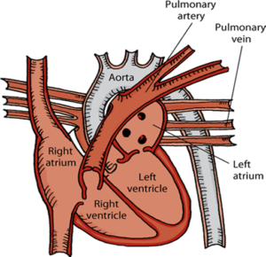
Fig. Cardiovascular system of horse
Examination of cardiovascular system
Examination of cardiovascular system of horse consist of following
- Physical examination,
- Echocardiogram,
- Electrocardiogram if an arrhythmia is present.
- Additional tests such as blood pressure measurement,
- Thoracic radiographs
- Laboratory tests that may be useful, depending on the problem, include CBC, serum chemistry panel, measurement of cardiac troponin, and blood cultures.
Physical examination
Physical examination includes the following
- Auscultation of heart
- Assessing mucous membranes for color and capillary refill time, pulse quality
- Examination of the extremities for warmth
- Notice whether there is generalized venous filling, edema, or a jugular pulse
- Normal heart rate – 28-48 bpm
LEFT SIDE
- Heart: Auscultation of the heart on the left cranial ventral thorax.
- A. M (Pulmonary, Aortic, Mitral)
- The Pulmonary valve – 3rd intercostal space at costochondral junction
- The Aortic valve – 4th intercostal space, just below the level of the shoulder
- The Mitral valve – 4th intercostal space at level of olecranon
RIGHT SIDE
- The Tricuspid valve – 3rd to 4th intercostal space between the olecranon and the costochondral junction
The 4 normal heart sounds that can be heard in the horse are
- S1 (atrioventricular valve closure)
- S2 (semilunar valve closure)
- S3 (rapid ventricular filling)
- S4 (atrial contraction)
- S1 and S2 are the “lub” and the “dub”.
- When all four heart sounds are heard, the sequence is S4, S1, S2, S3, pause for diastole, S4, S1, S2, S3, pause for diastole, etc. However, you may also hear S4, S1, S2, pause…. or S1, S2, S3, pause….These may sound like “gallop” rhythms, but are not considered to be abnormal in the horse.
Cardiac murmur
Cardiac murmurs are common in horses. Around 60 % of horses have murmurs, however, most are functional murmurs which are unrelated to heart disease especially when listen right after exercise or when the horse is excited. The critical part of the clinical examination is to identify those murmurs which are caused by cardiac disease, which may affect the athletic performance. Murmurs are generally characterized by location in the cardiac cycle (systolic vs diastolic), timing (early, mid, late or pan), intensity, and point of maximal intensity. The most common causes of pathologic systolic murmurs in horses include mitral or tricuspid valve regurgitation. By far the most common diastolic murmur in horses is caused by aortic valve regurgitation.

Arrhythmias
Arrhythmias are very common in horses and not always pathologic. In fact, the most commonly encountered arrhythmia, 2nd degree atrioventricular block, is most often caused by high resting vagal tone in relaxed, fit horses. It is also common to hear arrhythmias right after exercise, as the heart rate slows back down to its normal resting rate. This is thought to be caused by imbalances in sympathetic and parasympathetic tone, and the arrhythmia goes away once resting heart rate has returned. Other arrhythmias encountered in horses include atrial fibrillation and atrial or ventricular premature contractions. Electrocardiography is necessary to properly identify arrhythmias.
Electrocardiography
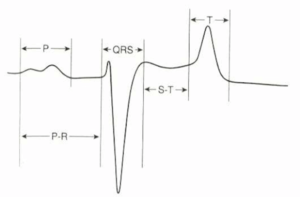
Electrocardiography records the electrical events of the cardiac cycle. In equine medicine, it is primarily important in the diagnosis of arrhythmias. Although it is possible to record the usual 6 leads in horses but “base-apex” system is commonly used. It consists of two electrodes, one positive and one negative, in a format called the base-apex lead. The positive electrode of lead I (left arm) is attached to the skin of the left thorax at the fifth intercostal space immediately caudal to the olecranon, and the negative electrode (right arm) is placed on the jugular furrow in the caudal third of the right neck
Serum cardiac troponin I concentration
The serum concentration of cardiac tropinin I provides an excellent cardiac biomarker in large animals, providing a sensitive and persistent indicator of cardiac injury. Healthy horses have cardiac troponin I concentrations below 0.11 ng/mL using the human immunoassay. Healthy neonatal foals have cardiac troponin I concentrations of less than 0.49 ng/mL.
Cardiovascular adaptations to exercise and training
- The functionally most important adaptation is the improvement in maximal cardiac output which is the result of an enlargement in cardiac dimension,
- An increase in blood volume, allowing for greater filling of the ventricles and a consequent larger stroke volume.
- The perfusion capacity of the muscle is increased, permitting for greater oxygen delivery.
- Decreased arterial resistance
- Adaptation in structure and number of artery , arterioles and capillary
- Exercise heart rates is six to seven times resting values
- Stroke volume – which is maintained by splenic contraction, increased venous return, and increased myocardial contractibility.
- Spleen contain one-third of total red cell volume.
- Despite the great changes in cardiac output, increases in blood pressure during exercise are maintained within relatively smaller limits, as both pulmonary and systemic vascular resistance to blood flow is reduced.