

FETAL ANASARCA AS A CAUSE OF DYSTOCIA: A CLINICAL REPORT
Nancy Jasrotia*, Nagaraj, Anju Kujur, Rohit Kurhe
*Corresponding author. e-mail: nancyjas53@gmail.com
ABSTRACT: A full term crossbred doe, aged 3 years and weighing around 35kg was presented in OPD VGO, Veterinary College, Bidar (KVAFSU, Bidar) with the anamnesis stating animal’s inability to deliver the fetus, persistent straining accompanied by restlessness and anorexia. On per vaginal examination, fetal limbs were palpable in the anterior vagina. After careful manipulation with the lubricated gloved hand per-vaginally and application of mild traction, single live but weak female fetus with anasarca was extracted out that died few mintues after birth. The doe recovered uneventfully after subsequent treatment with uterine cleanser and systemic antimicrobial therapy for 4 days. In conclusion, a case of dystocia due to fetal anasarca and its successful management is reported below.
Key words: Fetal anasarca, Goat, Dystocia, Per-vaginal delivery, Manual extraction
The fetal causes of dystocia in the goat are fetal maldisposition and fetal oversize. Fetal oversize occurs due to different dropsical conditions of the fetus such as fetal anasarca, ascites, edema of the allantochorion, hydrops of the amnion or allantois or both etc. Foetal anasarca is generalized edema of subcutaneous tissue of fetus which may either occur alone or along with other anomalies (Roberts, 1971). The condition is seen commonly in cattle but may also affect sheep, (Roberts, 1986) and goat (Tamuli et al., 1987; Sharma et al., 2002; Purohit et al., 2006). The definite cause of this is unknown and could be associated with achondroplasia or circulatory disturbance which occurs as a result of lymphoid tissue agenesis possibly due to existence of an autosomal recessive gene defect (Long, 1996; Monteagudo et al., 2002; Roberts, 1971). Most anasarcous fetuses are expelled dead but some may survive for few minutes after delivery. When there is difficulty in delivery of such fetus, cuts must be given over fetal surface to release the fluid or fetotomy and/or forced extraction may be carried out to deliver the fetus. In rare and complicated cases when vaginal delivery is not possible due to extreme oversize, caesarean section is indicated. If the condition is diagnosed before parturition by ultrasonography, therapy suggested is termination of the pregnancy. The following is a case study of dystocia due to fetal anasarca and its successful management in a crossbred goat.
CASE HISTORY AND OBSERVATION
A 3-year-old pluriparous, crossbred doe was presented at OPD (VGO), Veterinary College, Bidar, with rectal temperature of 100.30 F and history of completed gestation, persistent straining, restlessness, anorexia accompanied by animal’s inability to deliver the fetus. The animal was active with a good body condition score (Fig.1). The vulva of the animal was swollen and per vaginal examination done with a lubricated gloved hand, revealed two fetal forelimbs along with a fluctuating swollen head palpable in the anterior vagina.
DIAGNOSIS AND TREATMENT
Per vaginal examination revealed anterior longitudinal presentation of the fetus with both forelimbs and a fluctuating swollen head extended in the birth canal. After careful manipulation with the lubricated gloved hand per-vaginally and application of mild traction to the fetal limbs by holding them between thumb, forefinger and middle finger, single live but weak female fetus with anasarca was extracted out that died few mintues after birth (Fig.2&3). Further per vaginal examination and radiography confirmed the absence of any other fetal structures in the uterus.
After the delivery of anasarcous fetus, uterine douching was performed with povidone iodine solution. The animal was treated with antimicrobial (Enrofloxacin @ 5mg/kg. b.wt. IM OD) and other supportive therapy using Meloxicam @ 0.05mg/kg. b.wt. IM OD for 4 days along with Involon® (Indigenous herbal uterine cleanser) @ 100 ml PO as a loading dose followed by 50 ml for 4 days. The animal recovered uneventfully after 4 days of treatment.
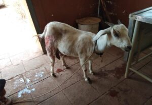
Figure 1: A non-descript doe with complete gestation period presented at OPD-VGO |
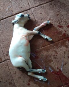 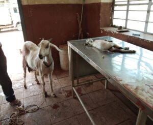
Figure 2 & 3: Anasarcous fetus delivered per vaginally by gentle traction |
DISCUSSION
Fetal anasarca, a type of fetal dropsy is of obstetrical importance as it generally results in dystocia due to increased diameter of the fetus. The condition occurs due to simple autosomal recessive gene (Roberts, 1971); is occasionally observed in kids, lambs and foals but is common in calves (Craig, 2000). Fetal anasarca or excessive edema of the fetus may develop in a single fetus or one of the twins (Roberts, 1971). The affected anasarcous fetus is usually carried to term, and concern is caused when there is lack of progress in second-stage labour which is due to the great increase in fetal volume caused by the excess of fluid in the subcutaneous tissues, particularly of the head and hind limbs (Noakes et al., 2001). The present case was also of single fetus pregnancy which was carried full term but resulted in dystocia due to fetal oversize. The delivered fetus was having generalized edema of the head and body with marked fluid accumulation in the peritoneal and pleural cavity and weighed 4.2kg. Various cases of fetal anasarca alone or in association with other developmental anomalies have been reported in goats (Chandrasekaran et al., 2015; Prabaharan et al., 2016; Dewry et al., 2019; Katiyar et al., 2020;
In removing large anasarcous fetuses, forced extraction is usually successful. If the fetus is too large, fetotomy and evisceration may occasionally be necessary. This is easily performed because the fetal tissues are soft, friable and edematous (Roberts, 1971). Most anasarcous fetuses are expelled dead while some may survive for few minutes after delivery. In the present case, extraction of the fetus was carried out with gentle traction per vaginally and the fetus was delivered live, however it died few minutes after birth. Caesarean section is indicated when per-vaginum delivery is not possible due to extreme fetal oversize or when fetal anasarca is coupled with other monstrosities like achondroplasia (Dewry et al., 2019)
SUMMARY
The clinical case of fetal anasarca as a cause of dystocia is not so common in goat. The management can be done successfully by forced extraction and/or fetotomy or caesarean section; the choice of which depends on the fetal size and severity of the condition.
ACKNOWLEDGEMENT
The authors are thankful to the faculty of Department of Veterinary Gynaecology and Obstetrics, Veterinary College, Bidar, KVAFSU, Karnataka for providing necessary facilities and guidance throughout the study of clinical cases.
REFERENCES
Chandrasekaran D, Selvakumar S, Suresh Kumar R, P. Pothiappan AKD, Balasubramanian S (2015) Per-vaginal delivery of anasarcous foetus in a tellicherry doe. Indian Journal of Animal Reproduction. 36 (1): 60-61.
Craig JF (2000) Flemings’s Vet. Obstetrics, Green World Publishers, London, U.K
Dewry RK , Talukdar D , Hazarika SB , Saikia B, Yadav HP (2019) Dystocia due to fetal anasarca coupled with achondroplasia of one foetus in twin pregnancy of a goat. International Journal of Science, Environment& Technology 8(2): 388-390
Katiyar R, Kharayat N, Rautela R, Kumar A, Gautam D, Ghosh SK, Prasad JK (2020) A Rare Case of Dystocia Due to Hydroamnion Coupled with Fetal Anasarca in a Doe. Agricultural Science Digest
Long S (1996) Abnormal development of the concepts and its consequences in: Veterinary Reproduction and Obstetrics, 7th Edition, W.B. Saunders Co Ltd.
Monteagudo L, Lujan L, Tejedor T, Climent S, Acin C, Navarro A, Arruga, MV (2002) Fetal anasarca (Hydrops foetalis) associated with lymphoid tissue agenesis possibly due to an autosomal recessive gene defect in sheep. Theriogenology, 58: 1219-1228.
Noakes DE, Parkinson TJ, England GCW (2001) Veterinary Reproduction and Obstetrics, 8th Edn., W.B. Saunders, London.
Prabaharan V, Sivakumar A, Jayaganthan P, Raja S, Vijayarajan A, Satheshkumar S (2016) Dystocia due to fetal anasarca and ascities with live fetus in a doe. Int. J. Sci. Environm. Tech. 5: 2586-2589.
Purohit GN, Gupta AK, Gaur M, Sharma A, Bihani DK (2006) Periparturient disorders in goats- a retrospective analysis of 324 cases. Dairy Goat Journal 84(2):24-33
Roberts SJ (1971) Veterinary obstetrics and genital diseases, (2nd edn).CBS Publishers and Distributors.
Roberts SJ (1986) Veterinary obstetrics and genital diseases, (3rd edn).CBS Publishers and Distributors.
Sharma SS, Purohit GN, Bishnoi BL, Yadav RC (2002) Fetal anasarca in a goat. A case report. Veterinary Practitioner 3: 47
https://www.pashudhanpraharee.com/cesarean-section-in-cow-buffalo/
Tamuli MK, Rajkonwar CK, Borgohain BN (1987) Foetal anasarca in a kid. A cause of dystocia. Indian Journal of Animal Reproduction 8(1): 63.