


Management of Congenital and Inherited Cardiovascular System Disorder in Horses
Bhand Akshata Chandrakant
PhD Scholar, Division of Medicine, Indian Veterinary Research Institute, Izatanagar, Bareilly – 243122. Email ID – drakshatavmc@gmail.com
Abstract
The cardiovascular system in horses serves as a vital network for blood transport and is comprised of blood vessels and a muscular pump, the heart. This system facilitates the transportation of essential substances such as water, oxygen, carbon dioxide, energy fuels, electrolytes, hormones, and metabolic products. Congenital and inherited disorders of the cardiovascular system in horses can arise from genetic defects, environmental factors, infections, pharmaceutical toxicity, maternal medication use, inadequate maternal nutrition, or a combination of these factors. Arabian horses exhibit a higher incidence of congenital defects, although the overall prevalence in the equine population remains low. Common congenital disorders include ventricular septal defect, patent ductus arteriosus, tetralogy of Fallot, and tricuspid dysplasia. Additionally, acquired heart and blood vessel disorders in horses are explored, focusing on degenerative valve disease, myocarditis, infective endocarditis, and pericardial disease. Degenerative valve disease, most prevalent in older horses, involves thickening of heart valves, leading to turbulent blood flow and potentially heart failure. Myocarditis, characterized by inflammation of the heart muscle, can result from bacterial or viral infections, parasitic infestations, toxin ingestion, or mineral deficiencies. Infective endocarditis, an infection of the endocardium, primarily affects cardiac valves and necessitates prolonged antibiotic treatment. Pericardial disease, encompassing pericarditis and cardiac tamponade, involves inflammation of the pericardium and fluid accumulation around the heart, leading to various clinical signs.
Keywords: Cardiovascular System, Patent ductus arteriosus, Ventricular septal defect, Tetralogy of fallot, pericarditis, etc.
The cardiovascular system is a blood-transporting network of blood vessels and a muscle pump that powers the heart. Water, oxygen, carbon dioxide, fuels for energy generation, electrolytes, hormones, and metabolic products are among the things it mostly transports (Marr and Bowen, 2011). Congenital and inherited disorders of the cardiovascular system in horses are as follows
Congenital and Inherited Disorders of the Cardiovascular System in Horses
They may be caused by genetic defects, environmental circumstances, infections, pharmaceutical toxicity, the dam’s use of the medication, inadequate nutrition for the mother, or a combination of these. High rate of incidence occur in Arabian horses and the overall number of horses with congenital defects is very low (Chaffin et al., 1992). The most common congenital defects in horses are
- Ventricular septal defect,
- Patent ductus arteriosus,
- Tetralogy of Fallot,
- Tricuspid dysplasia.
Ventricular septal defect
The ventricles, the heart’s two bottom chambers, are separated by a wall called the septum, which has an opening or defect known as a VSD. Normal development prevents oxygen-rich blood from mixing with oxygen-poor blood at birth by having the wall separating the chambers collapse before the fetus is born. Failure of the hole to close could result in decreased oxygen delivery to the body or increased heart pressure (Reef, 1995, Lange et al., 2021).

Figure 1: Ventricular septal defect
Signs
- A small defect usually causes minimal or no signs, and affected horses may be able to engage in moderate levels of physical activity.
- Larger defects may result in severe left-side congestive heart failure.
- The development of a right-to-left shunt is indicated by a cyanosis, fatigue, and exercise intolerance.
- Most affected animals have a loud murmur
Diagnosis – Chest x‑rays, echocardiography (ultrasonography), and other more specialized techniques may be used to confirm the defect.
Treatment – depends on the severity of signs, intended use of the horse, and direction of the shunt. Horses with small ventricular septal defects without complications do not typically require treatment and the outlook is good. Animals with a moderate to severe defect more commonly develop signs, and treatment should be considered. Treatment may include medications, surgery (uncommon), and the removal of excessive red blood cells, if present. Horses with a ventricular septal defect should not be bred.
Patent ductus arteriosus
The ductus arteriosus is a short, broad vessel in the unborn foal that connects the pulmonary artery with the aorta. It allows most of the blood to flow directly from the right ventricle to the aorta. In an unborn foal, oxygenated blood within the main pulmonary artery is forced into the descending aorta through the ductus arteriosus, bypassing the nonfunctional lungs. In most species, the ductus closes at birth when the animal begins to breathe, allowing blood to flow to the lungs. In foals, however, the complete closure of the ductus may be delayed for up to a week after birth, causing a heart murmur. These foals do not typically show any signs associated with the delayed closure (Dufourni et al., 2018).
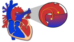
Figure 2: Patent Ductus Arteriosus
Tetralogy of fallot
It is caused by a combination of pulmonic stenosis (an obstruction of blood flow from the right ventricle), a ventricular septal defect, thickening of the muscle fibers of the right ventricle, and varying degrees of the aorta rotating to the right (Schmitz et al., 2008).
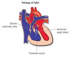
Figure 3: Tetralogy of Fallot
Signs
- Stunted growth, fatigue (especially after exercise), collapse, or seizures.
- A murmur is often, but not always, present.
Diagnosis: Electrocardiographs, x-rays, and echocardiography (ultrasonography) can help confirm the diagnosis.
Treatment – options include medications and periodic removal of excessive red blood cells (phlebotomy). The outlook is guarded to poor, but some foals with mild to moderate disease may reach adulthood.
Acquired Heart and Blood Vessel Disorders in Horses
- Degenerative valve disease
Degenerative valve disease is characterized by thickening of the heart valves. In horses, degenerative valve disease most often affects the aortic, mitral, and tricuspid valves. he mitral valve is most commonly affected. The condition generally occurs slowly over time and is most common in older horses. Failure of the valve to close properly turbulent blood flow Blood flows back into the previous chamber an increase in its blood volume and size. When regurgitation is severe, the chamber’s pressure may also increase. The body uses compensatory mechanisms to counteract the decreased blood flow. This results in further enlargement of the heart (Buergelt, 2003).
Signs
- The signs of failure vary depending on the affected valve.
- Severe mitral valve degeneration can lead to signs of left-side heart failure, such as trouble breathing or coughing.
- Severe tricuspid valve disease can lead to signs of right-side heart failure, such as fluid accumulation under the skin of the limbs and on the underside of the chest and abdomen.
- In many horses, signs of aortic valve degeneration are uncommon because the aortic regurgitation is rarely severe enough to cause heart failure.
Diagnosis: Diagnosis is made by physical examination, radiography and by Echocardiography.
- Myocarditis
Myocarditis is a local or widespread inflammation of the heart muscle, with degeneration or death of the heart muscle cells (Van Loon and Decloedt, 2012).
Etiology
- Bacteria – Streptococcus spp, Salmonella, Clostridium, and Borrelia burgdorferi.
- Viral – equine infectious anemia virus and equine influenza.
- Strongylosis (a parasitic infection) can also cause myocarditis.
- Less common causes include severe deficiencies of vitamin E or selenium
- Ingestion of cardiac toxins, blister beetles, and poisonous plants such as rubber vine or white snakeroot.
- Mineral deficiencies such as iron and copper.
Signs
Clinical signs of myocarditis include depression, lethargy, work intolerance, and increased resting heart rate. Arrhythmias are not uncommon. Clinical signs may be confused with mild colic.
Treatment – includes treatment of any underlying cause of the myocarditis (eg. antibiotics for a bacteremia, anthelmintics for parasites, vitamin E supplementation, etc.) and supportive care for the decreased cardiac function (anti-arrhythmic drugs, positive inotropes, etc.). Steroids are often given for their anti-inflammatory effects, especially if an arrhythmia persists. (Flunixin meglumine @1.1mg/kg B.wt SID for 5days)
Treatment of heart failure caused by valvular and myocardial disease
- Furosemide- 0.5-3.0 mg/kg q 12 hour (IV, IM, or PO) (note – oral absorption may be erratic, so parenteral administration is preferred)
- Digoxin- Loading dose (rarely used) 0.0044-0.0075 mg/kg IV q12h (2 doses only)
- Maintenance dose- IV:0.0022-0.00375 mg/kg q 12h; Oral:0.011-0.0175 mg/kg q12h (oral used more commonly)
- Signs of digoxin toxicity: anorexia, diarrhea, colic, weakness, depression, cardiac arrhythmias, and conduction disturbances.
Infective endocarditis
Infective endocarditis is an infection of the endocardium and typically involves one of the cardiac valves, although endocarditis of the cavity’s wall may occur (Henderson et al., 2020). Infection is caused by bacteria carried in the blood. The infection gradually destroys the valve and keeps it from working properly. Preexisting damage to the valve or immune dysfunction can increase the likelihood that infective endocarditis will occur. In horses, the aortic and mitral valves are most commonly affected. The tricuspid valve is rarely affected, and pulmonic valve infective endocarditis is exceedingly rare. Common isolates in equine bacterial endocarditis include Streptococcus zooepidemicus and Actinobacillus equuli.
Signs – Vary depending on where the infected blood clots lodge, but affected animals usually have a murmur, fever, weight loss, and fatigue. The condition can also lead to heart failure, which can result in trouble breathing (Buergelt et al., 1985).
Diagnosis- Diagnostic tests should include CBC, serum chemistry panel, blood culture, and echocardiography. CBC may show evidence of chronic infection (increased white blood cell count, increased fibrinogen, etc.). Serum chemistry panel will only show evidence of organic disease (eg. renal or hepatic) if the horse is in heart failure or if primary organ disease is the source of the bacteremia. While positive blood cultures are indicative of bacteremia, negative blood cultures do not rule out bacterial endocarditis; the infection may have cleared by the time the diagnostics are performed, yet the changes on the valves remain.
Treatment: Treatment of bacterial endocarditis includes long-term administration of antibiotics (preferably based on culture and sensitivity) and supportive care for cardiac dysfunction.
Pericardial disease
A. Pericarditis
Pericarditis is inflammation of the pericardium, which can lead to pericardial effusion. In horses, septic pericarditis (resulting from infection) and pericarditis of an unknown cause are the most commonly reported types of pericardial disease. Signs vary based on the amount of fluid that has accumulated (Perkins et al., 2004; Sprayberry et al., 2017).
- Common signs of pericarditis include:
- History of a recent respiratory tract infection
- Fever
- Lack of appetite
- Depression
- Less common signs of pericarditis include
- Weakness
- Distension of the jugular veins
- Muffled heart sounds
- Unwillingness or inability to exercise
- Listlessness
- Abdominal swelling due to fluid accumulation
B. Cardiac tamponade
The pericardium is the membrane that surrounds the heart. When fluid builds up within this membrane (called pericardial effusion), the heart is compressed. The pressure on the heart reduces its ability to pump blood. This condition is called cardiac tamponade. The compression significantly affects blood circulation and causes swollen jugular veins and accumulation of fluid in the abdomen. In addition, too little oxygen reaches the body’s tissues. Pericardial effusion rarely occurs in horses (Malalana et al., 2011).
Neoplastic disease
- The most common cardiac neoplasm is lymphosarcoma, which can invade the atria.
- Mesothelioma, melanoma, and hemangiosarcoma have been reported on the pericardium of horses, but these tumors are extremely rare.
Conclusion
In the realm of equine medicine, a thorough understanding of these cardiovascular disorders is imperative for veterinary professionals. Accurate diagnosis, effective management, and informed breeding practices are essential for ensuring the well-being of horses and maintaining their cardiovascular health. As ongoing research continues to shed light on these conditions, equine healthcare providers can refine their approaches to better address the complexities of cardiovascular disorders in horses.
References
Buergelt CD, Cooley AJ, Hines SA, Pipers FS. Endocarditis in six horses. Veterinary Pathology. 1985 Jul;22(4):333-7.
Buergelt CD. Equine cardiovascular pathology: an overview. Animal Health Research Reviews. 2003 Dec;4(2):109-29.
Chaffin MK, Miller MW, Morris EL. Double outlet right ventricle and other associated congenital cardiac anomalies in an American miniature horse foal. Equine Veterinary Journal. 1992 Sep;24(5):402-6.
De Lange L, Vera L, Decloedt A, Van Steenkiste G, Vernemmen I, van Loon G. Prevalence and characteristics of ventricular septal defects in a non‐racehorse equine population (2008‐2019). Journal of Veterinary Internal Medicine. 2021 May;35(3):1573-81.
Dufourni A, Decloedt A, De Clercq D, Saey V, Chiers K, van Loon G. Reversed patent ductus arteriosus and multiple congenital malformations in an 8‐day‐old Arabo‐Friesian foal. Equine Veterinary Education. 2018 Jun;30(6):315-21.
Henderson B, Diaz M, Martins C, Kenney D, Baird JD, Arroyo LG. Valvular endocarditis in the horse: 20 cases (1993–2020). The Canadian Veterinary Journal. 2020 Dec;61(12):1290.
Malalana F, Bardell D, McKane S. Idiopathic aseptic pericardial effusion with cardiac tamponade in a horse. Equine Veterinary Education. 2011 Feb;23(2):64-8.
Marr C, Bowen M, editors. Cardiology of the Horse. Elsevier Health Sciences; 2011 Jan 7.
Perkins SL, Magdesian KG, Thomas WP, Spier SJ. Pericarditis and pleuritis caused by Corynebacterium pseudotuberculosis in a horse. Journal of the American Veterinary Medical Association. 2004 Apr 1;224(7):1133-8.
REEF VB. Evaluation of ventricular septal defects in horses using two‐dimensional and Doppler echocardiography. Equine Veterinary Journal. 1995 Sep;27(S19):86-95.
Schmitz RR, Klaus C, Grabner A. Detailed echocardiographic findings in a newborn foal with tetralogy of Fallot. Equine Veterinary Education. 2008 Jun;20(6):298-303.
Sprayberry KA, Slovis NM. Sales performance and athletic outcome in young Thoroughbreds with pericarditis. Equine veterinary journal. 2017 Nov; 49(6):729-33.
Van Loon G, Decloedt A. Inflammatory myocardial disease and toxicity in horses: a recent update. InVeterinary Cardiovascular Society Autumn meeting 2012.
Minimizing Congestive Heart Failure in Dogs: Methods for Preventing and Management