


MANAGEMENT OF POST–CERVICAL UTERINE TORSION IN A MURRAH BUFFALO: A CASE REPORT
Kailash Kumar*, Archana Choudhary, Dushyant Dev Bhal, Surendra Patel and Satish
Vcterinary Clinical Complex
Arawali Veterinary College, Sikar-332403, Rajasthan, India
*Corresponding author Email: kailashkumar7006@gmail.com
Abstract
Four-year-old full term pregnant murrah buffalo was presented with history of restlessness, frequent lying down and getting up, anorexia and excessive straining with no progress of parturition from last 6 hours. On per vaginal examination, the clock wise (to the cow’s right) post cervical uterine torsion was diagnosed along with the help of anamnesis. After stabilization of the animal, the uterine torsion was amended by Schaffer’s method and the live foetus was relieved by simple traction.
Keywords: Uterine torsion, buffalo, modified Schaffer’s method.
Introduction
Uterine torsion is defined as the twisting of the uterus on its longitudinal axis and usually occurs in a pregnant uterine horn (Purohit et al., 2011a, b). The pregnant uterus rotates about its long axis, with the point of torsion being the anterior vagina just caudal to the cervix (Purohit et al., 2011a, b) (post-cervical uterine torsion). Less commonly the point of torsion is cranial to the cervix (pre-cervical uterine torsion). It is observed commonly in multiparous animals at the time of parturition or during the last month of gestation (Satish and Gaur, 2019). Uterine torsion is the most frequent cause of dystocia in buffalo, followed by incomplete dilatation of cervix and uterine inertia (Satish et al., 2022). In the present communication, successfully management of a case of dystocia due to uterine torsion in murrah buffalo is reported.
History and Observations
A four-year-old murrah buffalo in its 1st parity was brought at 11 pm to the Veterinary Clinical Complex, Arawali Veterinary College, Sikar with a history of completion of gestation period, colic, restlessness, frequent sitting down and getting up, tail twitching, anorexia and non progressive labour with intermittent straining from last 6 hours. Animal was previously handled by a quack. On general clinical examination, the animal appeared dull and reluctant to move; the rectal temperature was 100.7℉ and the pulse and respiratory rates were normal. Per rectal examination of the animal indicated more than 180° right-sided uterine torsion. Per-vaginal examination revealed a strong twist in the anterior vagina running towards the right side and which brings the confirmative diagnosis of post cervical clockwise (to the cow’s right) uterine torsion.
Treatment and Discussion
After confirmation of the uterine torsion fluid therapy with rintose 20% (2 liter) was administered along with broad spectrum antibiotic ceftriaxone (10 mg/kg body weight) was administered parenterally to combat toxemia and shock before handling of case.
After the treatment, the animal was cast on right lateral recumbency (on the same side of torsion) with both the fore limbs and hind limbs tied distinctly. The wooden plank was placed in an oblique direction over the flank region without any bone involvements in order to fix the uterus externally. Then the animal was slowly rolled towards the same side of torsion (right). Per-vaginal examination following every complete rotation was done. Immediately after two successful rotations, the intact amniotic sac came out and the dilated cervix with a fetus could be palpated. After manual rupturing of the amniotic sac a live calf anterior longitudinal presentation (P1), dorso-sacral position (P2) with extended forelimbs (P3) was relieved by gentle traction. The foetal membrane was shed after 3 hours of successful relief.
After removal of the fetus, the animal was treated with inj. oxytocin (30 IU intramuscularly) and 450 ml of calcium magnesium borogluconate (Mifex®) were administered slow intravenously as well as nitrofurazone and urea pessary was placed inside the uterus to avert infection. Liquid Utrevive® (100 ml orally twice a day) for five days was given.
Uterine torsion accounts for most of the cases of maternal dystocia in this species, and the direction is to the right in more than 90% of the cases (Satish and Gaur, 2019). Uterine torsion is considered to be a more frequent maternal cause of dystocia in buffaloes compared to cattle (Purohit et al., 2012). Predisposing factors include relatively long uterine ligaments, the low number of smooth muscle cells in the broad ligament, constant confinement and hilly terrain (Ghuman, 2010).
Modified Schaffer’s method (non-surgical) with abdominal pressure was found to be the most successful procedure for correction of uterine torsion in buffaloes and is safe and easy to handle.
References
Ghuman SPS (2010). Uterine torsion in bovines: a review. Indian Journal of Animal Sciences 80(4):289-305.
Purohit GN, Barolia Y, Shekher C and Kumar P (2011a). Diagnosis and correction of uterine torsion in cattle and buffaloes. Raksha Technical Review 2:11-17.
Purohit GN, Barolia Y, Shekher C and Kumar P (2011b). Maternal Dystocia in cows and buffaloes: A review. Open journal of Animal sciences 1(2):41-53.
Purohit GN, Kumar P, Solanki K, Shekher C and Yadav SP (2012). Perspectives of fetal dystocia in cattle and buffaloes. Veterinary Science Development 2:31-42.
Satish and Gaur M (2019). Studies on the Therapeutic Approaches for Uterine Torsion in Surti Buffaloes (Bubalus bubalis). The Indian Journal of Veterinary Sciences & Biotechnology 14(3):36-39.
Satish, Kumar D, Gaur M, Purohit GN, Jhamb D and Chahar SK (2022). Retrospective prevalence analysis of dystocia in buffaloes with special reference to uterine torsion. Ruminant Science 11(2):471-474.
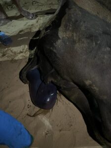
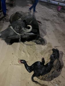
Fig.1. Intact amniotic sac came out Fig.2. Live fetus delivered after
immediately after detortion correction of torsion of uterus