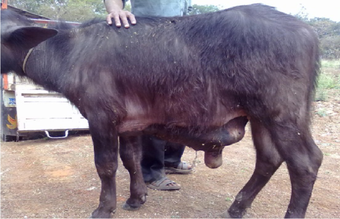
Obstructive Urolithiasis in Male Buffalo Calves: Treatment and Management
Urolithiasis is the lodgment of uroliths, anywhere in the urinary system but most frequently at the distal end of sigmoid flexure in ruminants that results in obstruction of urine flow . Urolithiasis is more common in male ruminants compared to females due to anatomical conformation of the urethral tract . The female has short, wide, and straight urethra, while the male has long, narrow and tortuous urethra which makes them more prone to urethral obstruction, particularly distal aspect of the sigmoid flexure in bovines and urethral process in sheep and goats. The gradual narrowing of the urethral orifice is a major predisposing factor for obstructive urolithiasis . In addition, factors such as diet, age, breed, genetic makeup, season, soil, water, mineral, and urinary tract infections plays an important role in the formation of urolithiasis . The clinical signs and physiological parameters of urolithiasis may vary with the degree of urethral obstruction, its duration, age and sex of the animals, and status of urinary bladder and urethra. Urethral obstruction in calves is a fatal disorder that predisposes to high mortality rate unless the animal is subjected to emergency surgical treatment for correction of the obstruction.
The incidence of retention of urine is more in male bovines as compared to female bovines followed by canine, equine, caprine and ovine. Retention of urine can be due to obstructive urolithiasis, urethral stenosis, urethral fistula and testicular tumour in bovines and equine. However, the most common etiological factor for retention of urine in male bovine calf is obstructive urolithiasis and this condition is generally encountered more in winter season. Urolithiasis in ruminants is of considerable economic importance as it results in heavy losses to the farmers.
ETIOLOGY OF OBSTRUCTIVE UROLITHIASIS
The condition is multifactorial and some of the possible causes are as under · The formation of calculi depends upon the diet fed to the animals. The animals maintained on high concentrate diet usually develop phosphate calculi because concentrate are rich in phosphorus. These phosphate calculi are smooth soft small and multiple in number. · In the second category, the animals maintained on high roughage diet develops carbonates and silicious calculi because roughage is rich in silica. These calculi are rough hard white and usually single in number. · Urine pH also plays a major determining factor for calculus formation. Mostly urine of herbivore is alkaline and alkaline urine favours precipitation of calcium and magnesium salts. · Water intake also plays an important role in calculus formation. Less water intake during winter/water deprivation leads to calculus formation. · Other factors like vitamin A and vitamin D deficiency will cause desquamation of lining of urinary tract. Site of lodgement of calculi: The calculus formation begins its development in the pelvis of the kidney but majority of calculus are flushed out through the urine but if it hinders the urinary passage the signs of obstructive urolithiasis develops. The calculi usually get lodged at the site of sigmoid flexure in bovines, in the terminal portiont of urethra/ glans penis in camels, urethral process in sheep and goat and just caudal to ospenis in dogs.
CLINICAL SIGNS
The clinical signs depends on the degree of obstruction whether partial or complete. In partial obstruction of urethra there is dribbling of urine, dysuria, abdominal pain/colicky symptoms, haematuria (due to inflammatory conditions of urinary tract). In cases of complete obstruction there is teeth grinding, stamping of legs, constant straining that often leads to rectal prolapse. Due to obstruction in the outflow of urine, the urine gets accumulated in the kidneys and will cause dilation of entire urinary tract ,the enlarged kidney due to storage of urine is known as hydronephrotic kidney and if the urine gets infected the condition is known as pyelonephrotic kidney. If the obstruction is not removed after a gap of 48 to 75 hours then urinary bladder/urethra will get ruptured and urine gets lodged in abdominal cavity which leads to bilateral distention of abdomen known as water belly abdomen. Clinical signs of rupture of urinary bladder are salivation, dehydration and sunken eyes. Urethral rupture will lead to subcutaneous infiltration of urine and with due course of time it will lead to formation of urine scald. Due to accumulation of urine in abdominal cavity /peritoneal cavity there will be diffusion of electrolyte from serum /blood towards abdomen resulting in hyponatremia, hypokalemia, hypochloremia, hypocalcemia. Due to accumulation of sodium (Na+) and chlorine (Cl-) in abdominal cavity, water from intracellular and extra cellular space will move to abdominal cavity and a state of dehydration occurs. Abnormal concentration of urea, creatinine and other nonnitrogeneous waste products results into a stage of azotaemia and this may be due to prerenal/renal/postrenal involvement. Due to obstructive urolithiasis there will be metabolic alkalosis in ruminants which further alters the renal functioning and results in uremia (always postrenal).
DIAGNOSIS
- On the basis of history and clinical signs · Abdominocentesis · Radiography · Ultrasonography · Serum biochemistry by estimating sodium, potassium, chloride, calcium, phosphorus, total protein, creatinine and blood urea nitrogen.
TREATMENT
The first line of treatment involves evacuatiuon of urinary bladder which in large animals is done through rectal approach and in small animals by abdominocentesis. Then the calculus is removed by means of urethrotomy either by post scrotal approach or by performing ischial urethrotomy in the first phase calculi is removed and in the second phase bladder is repaired by performing cystorrhaphy Apart from these in small calf tube cystostomy can be done in which a incision is given in the paramedian region between the two rudimentary teats under local anaesthesia. After incising the muscles and peritoneum the bladder is approached. A second incision is given on the skin and a tunnel is created through which the catheter (Foley’s rubber catheter or silicon catheter) is passed and placed in the bladder. The muscles and the peritoneum are sutured with absorbable suture material (chromic catgut No. 1) and skin is sutured with non absorbable silk No. 1. The owner is advised to feed the calf with ammonium chloride at a rate of 0.5-1 gram per kg body weight and to restrict the flow of urine from the catheter using cap of a disposable syringe. Ammonium chloride is a urinary acidifier and thus helps in dissolution of the calculi and restriction in the flow of urine makes the animal to strain and with due course of time as a combined effect of dissolution of calculi and straining of animal the obstruction is relieved.
POST-OPERATIVE TREATMENT
TREATMENT
Intravenous fluid (NSS) For 3-5 days– To check dehydration
Acid/vinegar water is used —-To combat metabolic alkalosis
Inj. Neostigmine @ 2-4 mg by subcutaneous route 2-3 times for 3 days.—- For atony of urinary bladder
Systemic antibiotics intramuscular for 3-5 days— To check retrograde infection
Ammonium chloride @ 0.5-1 gm per kg body weight (till normal micturition)——- Urinary acidifier
NSAIDs intramuscular for 5 days—— To check pain.
Compiled & Shared by- This paper is a compilation of groupwork provided by the Team, LITD (Livestock Institute of Training & Development)
Image-Courtesy-Google
Reference-On Request


