


Successful Surgical Excision and Management of Carpal Hygroma in a Bullock
Dr. Ashwani Kumar Bassan
Veterinary Assistant Surgeon , Veterinary Dispensary Rathana ,Department of Animal Husbandry ,Distt. Jammu. 181111(UT, J&K)
Abstract
The report describes a case of unilateral carpal hygroma of right forelimb in a 3 years old HF CB bullock, successfully managed by surgical excision of fibrous mass without exposing the joint capsule under IVRA followed by pressure bandaging and the animal recovered uneventfully within 15 days.
Key words: bullock, hygroma, carpal joint, bursa.
Introduction
Carpal hygroma is a fluid-filled localized swelling that develop over the dorsal aspect of carpal joint involving the skin, a small subcutaneous precarpal bursa and loose subcutaneous connective tissue. The main predisposing factors are repeated trauma due to lack of bedding on hard floors, poorly designed manger and narrow or short stalls, which restrict free movement of the animal (Tyagi and Singh, 2006). Some hygromas are congenital while others are develop over time, usually in response to trauma (Venugopalan,2009). The most common cause of bursitis is direct trauma. Severe trauma leads to acute bursitis, whereas continuous and repeated trauma leads to chronic bursitis. Sometimes, the bacterial infection and toxemia also results in development of bursitis (Fathy and Radad 2006). The present paper reports successful surgical excision and management of carpal hygroma in a bullock.
Case History and Observations
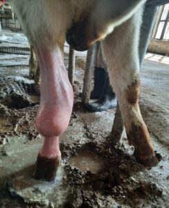
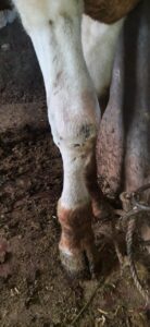
A three year old HF cross-bred bullock was presented with the history of non-painful, soft swelling over the dorsal aspect of carpal joint since last 2 months. It was previously treated by paravet, but animal doesn’t respond to the treatment and it grows steadily over a period of time (Fig. 1). On physical examination, it was found that the swelling was hard cranially and soft caudally i.e. towards carpal joint. On clinical examination mucous membrane was found to be slightly pale and temperature and other physical parameters were within normal range. Aspiration is done at most distal end of swelling revealed straw color fluid. The case was diagnosed as hygroma.
- Veterinary Assistant Surgeon in Animal Husbandry Department, UT, J&K and corresponding author
E mail : drashwanivet87@gmail.com.
Treatment
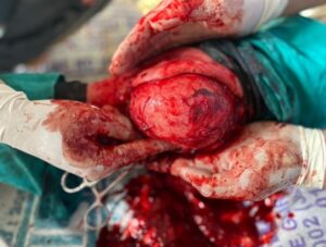
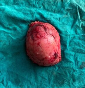
Animal was sedated by giving inj. Xylazine @ 0.025mg/kg b.wt intravenously. After that animal was restrained in left lateral recumbency. Pre-operative inj. Intacef Tazo 333.75mg and inj. maxxtol @0.4mg/kg was given intravenously. Under mild sedation tourniquet was applied proximal to the carpal joint and distal to the elbow joint and intravenous regional anaesthesia (IVRA) was achieved by giving 20 ml of the 2% lignocaine intravenously after removing 20 ml of blood from the cephalic vein. Surgical site was scrubbed for aseptic excision. Under local infilteration of 2% lignocaine, 3 cm long stab incision was given at anterior-lateral part of the swelling with sufficient allowance for skin closure without any tension. Skin was reflected by blunt dissection and the whole fibrous soft tissue mass (bursa) (Fig. 2) was surgically excised without exposing the joint (Fig. 3). The entire cavity was separated from the knee joint without opening the knee joint. The cavity was then packed with sterile gauge for preventing contamination during surgery and flushed with aqueous solution of normal saline mixed with betadine. Subcutaneous tissue was apposed using chromic catgut No. 2 in simple continuous pattern to favour adhesion formation and to obliterate the dead space. Skin was sutured with Silk No. 2 using horizontal mattress suture pattern and loosing the tourniquet in the meantime. Firstly the the entire carpal joint ( suture line) was cleaned with betadine then immobilized with thick layer of cotton and primary bandage and finally compression bandage (Nawar) to avoid the pressure on the suture line (Fig.4).
Post –operative care and management
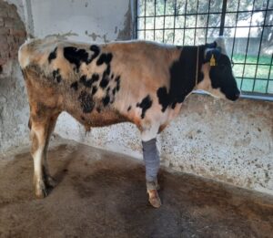
Post-operative care was done with antibiotics inj. Intacef tazo 3375mg iv daily for 5 days and analgesic inj. Maxxtol @ 0.4mg/kg Once Daily for 3days and tablet Melonex Zplus 2tab bid x 5 days. Owner was advised to keep the animal in stall confinement and applied compression bandage for 15 days. Second bandaging was done on 7th day with0.1% Povidone iodine solution revealed no complications with stable suture line and animal started bearing weight completely on the operated limb within a week. The skin sutures were removed after 15 days showed uneventful recovery without any post-operative complication ( Fig. 5). Animal was follow up upto 45 days postoperatively.
Discussion
In the present case hygroma was develop due to maintaince of the animal on hard floor and due to repeated trauma over the knee, skin gets thickened and a mass of thickened fibrous tissue developed beneath the skin ( Chhatpar et al., 2012). So due to chronic nature of hygroma, surgical excision resulted in uneventfull recovery. The surgical excision was successful in 16 out of 17 cases of precarpal bursitis in cattle (Piguet et al., 1997). The IVRA technique for local anaesthesia in patients undergoing surgical excision of carpal hygroma is very rewarding and easy to apply. The immobilization of the carpal joint with compression bandaging is very benficial for the stability of suture line and animal start weight bearing completely on the operated limb within a week with no post-operative complication (Shukla et al., 2020).The recovery in such cases has been reported to be successful (Lemay, et al., 2000; Chhatpar, et al., 2012; Piguet, et al., 1997).
Conclusion
Surgical excision is useful to treat the carpal hygroma in chronic cases (Chhatpar, et al., 2013). Proper post- operative care, good management and maximum rest provides uneventful recovery within 20 days without any post-operative complication.
Successful Surgical Excision and Management of Carpal Hygroma in a Buffalo
References
Chhatpar, K.D., Jora, G.K. and chuclasana, P.J. (2012). Carpal hygroma and its surgical excision in a cow . Intas Polovet, 13 (11): 279-280.
Fathy, A., and Radad, K. (2006). Surgical Treatment and Histopathology of Different Forms of Olecranon and Presternal Bursitis in Cattle and Buffalo. Journal of Veterinary Science. 7(3):287-91.
Lemay, A., Couture, Y., and Babkine,M. (2000). Surgical Treatment of Carpal and Tarsal Hygromas of The Dairy Cow: Review Of 11 Cases (1992-1998). Médecin Vétérinaire du Québec. 30(3):173-4.
Piguet, M., Steiner, A., Eicher, R., and Martig, J.(1997). Surgical Treatment of Carpal Hygroma in Cattle: 17 Cases (1990-1994).SchweizerArchiv fur Tierheilkunde. 139(5):210-6.
Tyagi, R.P.S., and Singh, J. (2006). Ruminant Surgery, 1stEdn, CBS Publisher and Distributors, New Delhi. pp. 312-313.
Venegopalan, A. ( 2009). Essentails of veterinary surgery ( 8th Ed.), Surgical Conditions Affecting Bursae, Capped Knee, Pg 155
|
|
|
|||
|
||||
|
|
|