SURGICAL MANAGEMNET OF SERTOLI CELL TUMUOR IN A CRYPTORCHID DOG – A CASE REPORT
Dr. M. Vigneswari
Shelter Veterinarian
People for Animal
Ponda, Goa, India – 403401.
ABSTRACT
A 8 year old male Labrador Retriever was presented to People for Animal, Ponda, Goa with the history of ventral abdominal distension for past 2 months with reduced appetite, dullness and constipation. On physical examination, the dog showed signs of enlarged mammary glands, an unusual odor and cryptorchid testis (right testis). Haematobiochemical values were within normal range except reduced hemoglobin count. Surgical correction was carried out and the mass was sent for histopathological examination and confirmed the case as sertoli cell tumor. Postoperatively, antibiotics and analgesia were administered and animal made an uneventful recovery.
Key words: Sertoli cell tumor, feminization, dog.
INTRODUCTION
Testicular tumors involving sertoli cell and germinal cells occur less frequently in comparision to interstitial cell tumors and are encountered rarely in extra testicular site (Doxsee et al. 2006). About one third of sertoli cell tumors (SCT) are hormonally active causing signs of endocrine imbalance like hyperestrogenism, feminization, gynecomastia, alopecia, bone marrow suppression and atrophy of contra lateral testicle (Kennedy et al. 1998). The association of SCT with cryptorchidism is well established (Post and Kilburn, 1987). The present paper describes about the diagnosis and surgical management of sertoli cell tumour in a cryptorchid testis in a Labrador Retriever dog.
Case history and Observation
A 8 year old male Labrador Retriever was presented to People for Animal, Ponda, Goa with the history of ventral abdominal distension for past 2 months with reduced appetite, dullness and constipation. On physical examination, the dog showed signs of enlarged mammary glands, an unusual odor and cryptorchid testis (right testis) (Fig 1). Haematobiochemical values were within normal range except reduced hemoglobin count. Based on the history and clinical signs, the case was diagnosed as cryptorchid testes with tumour. Surgical correction was carried out under general anaesthesia.
Surgical procedure
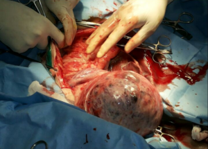
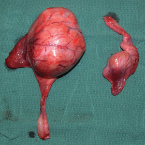
The animal was anesthetized with intravenous inj. Diazepam @ 0.5mg/kg and Inj. Propofol @ 3mg/kg IV. Under aseptic condition, the inguinal region was shaved and prepared for surgery. Incision was made directly on the skin over the tumor mass at the inguinal area. After skin and subcutaneous tissue incisions, blunt dissection was used to expose and free the tumor testicle from underlying tissues. Following orchidectomy, the left testis was greatly increased in size, presented multiple spermatic cord torsions (Fig 2) and was weighing about 750gm and measuring about 16 × 12 cm in length and had a significant profile alteration with multilobular appearance; cross-sectionally, it appeared completely replaced by spongy neoplastic tissue. The right testis was of normal size and not neoplastic (Fig 3). Tumour mass was sent for histopathological examination and the case was confirmed as sertoli cell tumour showing tubules as well as small nests of Sertoli cells in a hyalinised stroma (Fig 4). Postoperatively, oral cefpodoxime @ 10mg/kg for 7 days and meloxicam @ 0.2mg /kg for 3 days and sutures were removed on 10th postoperative day and animal made an eventful recovery.
RESULTS AND DISCUSSION
Sertoli cell tumors, interstitial cell tumors, and seminomas are the three common testicular tumour in dogs. Sertoli cell tumors were the most common type in cryptorchid canine testicle in the present study and this is in accordance with the findings of Liao et al. 2009. This difference may be explained by the limited cases of cryptorchid testicular tumors reported. The clinical signs of our study like presence of one hypertrophied testis along with atrophied testicles, feminization syndrome, gynecomastia were in accordance with the findings of Mischke et al. 2002. Cryptorchid testicles are generally intra-abdominal (within the abdomen), but may be under the skin in the inguinal area, or the area of the body where the hind leg meets the body wall. Bilateral orchiectomy is the treatment of choice for sertoli cell tumors. This is because about 50% of dogs diagnosed of this condition have bilateral tumors but only 12% are clinically detectable in the contralateral testicle (Lawrence and Saba, 2013).
Retained neoplastic testes are considered to be more predisposed to testicular torsion that can aggravate the clinical signs and result in guarded prognosis (Pearson and Kelly, 1975). In the cases reported above, spermatic cord torsion did not provoke congestive or necrotic changes although it certainly contributed to abdominal pain. This study emphasizes the importance of monitoring cryptorchid dogs, especially given the possible complications which may occur over time, in order to correctly evaluate the risks and benefits of surgical gonad removal.
REFERENCES
Doxsee, A.L., Yagar, S.J., Best, S.J. and Foster, R.A. (2006). Extratesticular intestinal and Sertoli cell tumors in previously neutered dogs and cats. A report of 17 cases. Can. Vet. J. 47:763-766.
Kennedy, P.C., Cullen, J.M., Edward, J.F., Goldschmidt, M.H., Larsen, S., Munson, L. and Nielson, S. (1998). Histological classification of genital system of domestic animals.Washington DC, Armed Forces Institute of Pathology.
Lawrence J.A and Saba, C.F. (2013). Tumors of the male Reproductive system. In: Withrow and MacEwen’s Small Animal Clinical Oncology. 5th Edn., Elsevier Saunders, USA.; p. 557–571.
Liao, A.T., Chu, P.Y., Yeh, L.S., Lin, C.T. and Liu, C.H. (2009) A 12-year retrospective study of canine testicular tumors. J. Vet. Med. Sci., 71: 919-923.
Mischke, R., Meurer, D., Hoppen, H.O., Ueberschar, S. and Hewick-Trautwein, M. (2002). Blood plasma concentration of oestradiol-17 beta, testosterone and testosterone/oestradiol ratio in dogs with neoplastic and degenerative testicular diseases. Res.Vet. Sci.73:267-272.
Pearson H, Kelly DF. (1975). Testicular torsion in the dog: a review of 13 cases. Vet. Rec., 97: 200-204.
Post, K and Kilborn, S.H. (1987). Canine Sertoli cell tumors: A medical record search and literature review. Can. Vet. J. 28:427-431.
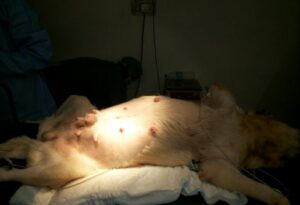
Fig 1: Gynecomastia in a male dog with a Sertoli cell tumor. |
Fig 2: Sertoli cell tumor of a cryptorchid testicle with torsion
|
|
Fig 3: Testicle (left) after surgery. Note the size difference between the normal testicle (right) and testicle with the tumor (left).
|
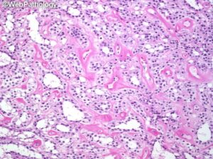
Fig 4: Sertoli cell tumour showing tubules as well as small nests of Sertoli cells in a hyalinised stroma.
|