

Swine Erysipelas
Lalrinkima1*, R. Ralte2, S. Azmi3
1Assistant Professor, Department of Veterinary Pathology, Khalsa College of Veterinary and Animal Sciences
2Associate Professor, Department of Veterinary Microbiology, Khalsa College of Veterinary and Animal Sciences
3Professor & HOD, Department of Veterinary Pathology, Khalsa College of Veterinary and Animal Sciences
Introduction
Swine erysipelas is an infectious disease caused by Erysipelothrix rhusiopathiae seen mainly in growing pigs. The diseases is also known as diamond skin diseases. The disease may be acute, sub-acute or chronic form. Clinical manifestations of the diseases are cutaneous erythema including characteristic diamond-shaped lesions, septicemia, arthritis and endocarditis. Erysipelas is a common cause of carcass condemnation at abattoirs.
Etiology
Erysipelothrix rhusiopathiae is the causative agent of swine erysipelas. The organism is gram positive, pleomorphic, non-spore forming, non-motile, encapsulated, facultative anaerobic, small rods (smooth form) or filaments (rough form) and can grow on non-enriched media.
Transmission
Swine is the most important reservoir host and pigs can carry the organism in the oropharynx, the organism can be cultured from the tonsils of clinically healthy pigs. An infected or sub-clinically diseased pig is often the source of infection to other herd animals. The bacterium is shed into the environment and susceptible pigs may acquire the infection by ingestion of contaminated soil or water (most common), percutaneous through skin wounds or possibly via ticks and flies.
Recovered pigs and those chronically infected might be carriers of the organism. The mode of entry is by ingestion and through skin abrasions. Following ingestion, the organism most likely enters the body via the tonsils or lymphoid tissue of the GI tract. In small populations kept in backyards, orchards or paddocks there is plenty of opportunity for access by birds and rodents and the persistence of the organism in soil favors persistence of disease.
Clinical signs
Clinical signs of swine erysipelas can be divided into acute, sub-acute and chronic forms. Subclinical infection can also occur where no disease is apparent, but may lead to chronic disease. The septicaemic and cutaneous (‘diamond’) forms are acute while arthritis and vegetative endocarditis are chronic forms of the disease.
Acute cases occur after an incubation period of 1-7 days with sudden death with high rise in temperature (104-106°F), stilly gait and get up with difficult suspended bowl material movement. Diamond skin lesions from sick pigs often have reddened or cyanotic skin, especially about the ears, snout, jowls, throat and ventral abdomen.
Chronic form is the most common form of erysipelas that may follow acute or sub-acute disease as well as subclinical infection and is characterized most commonly by arthritis. Arthritis mainly involved joints are hock, stifles, knee and elbow.
 |
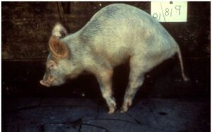 |
||
Figure 1: Typical diamond-shaped skin lesions. Figure 2: Arthritis in a growing pig.
Postmortem findings
Rhomboid (diamond skin) lesions are features of acute form of swine erysipelas and these lesions are pathognomonic for swine erysipelas. In pigs died due to acute form, it is often very visible presence of diffuse cutaneous hemostasis especially around the snout, ears, jowls, throat, abdomen and thighs. Lungs might showed congestion and edema. In addition to skin lesions, other lesions typical of septicemia are observed including enlargement and congestion in lymph nodes and spleen. Petechiae and ecchymoses may be seen in the renal cortex and heart (epicardium and atrial myocardium). Joints may enlarged and the synovium and periarticular tissues are typically distended by serofibrinous exudates that may also fill the joint cavity. Valvular endocarditis can be seen as proliferative, granular growth on the heart valves (mitral valve most common).
Diagnosis
Diagnosis is based on clinical signs, gross lesions and response to antimicrobial therapy and demonstration of the bacterium or DNA in tissues from affected animals. Isolation of E. rhusiopathiae from blood of affected pigs especially after enrichment is possible in acute cases and helps establish a diagnosis. In addition, molecular methods capable of detecting E. rhusiopathiae DNA in affected tissues or blood (PCR assays) can also be used. Recently, immuno-histochemical methods to demonstrate the organisms in formalin-fixed paraffin-embedded tissues have become available and are useful in cases when pigs have been treated with antimicrobials before sample submission.
References
- Department of Primary Industries. “Swine Erysipelas, Animal Biosecurity and Welfare Introduction.” (2017). (2nd edn). 1-2.
- Jeffrey J. Zimmerman et al. (2012). Swine diseases 10th edition, p 750-760.
- http://www.merckvetmanual.com/mvm/index.jsp?cfile=htm/bc/50902.htm
- Shimoji, Y, Ogawa Y, Tsukio M, and Shiraiwa K, et al. (2019). “Genome-wide Identification of Virulence Genes in Erysipelothrix rhusiopathiae: Use of a Mutant Deficient in a Tag Homolog as a Safe Oral Vaccine against Swine Erysipelas.” Infect Immun 87: 673-719.