



Canine Ehrlichiosis with Renal and Hepatobiliary Involvement in a Pomeranian Dog: A Case Report
Aishwarya G*1
1 Fourth professional year student, CVSc, Rajendranagar, P V Narsimha Rao Telangana Veterinary University, Hyderabad, 500030, Telangana, India.
Abstract
Sesha, a seven-year-old female Pomeranian dog, was presented to the clinic with a history of vomiting, inappetence, and progressive dullness. On presentation, the dog was dehydrated, depressed, and had pallor of visible mucous membranes. Hematological laboratory tests revealed anemia and severe leukocytosis. Serum biochemical analysis showed increased alkaline phosphatase, hyperbilirubinemia, hypoproteinemia, and hypoalbuminemia. Kidney function tests demonstrated severe azotemia with marked increase in urea, blood urea nitrogen, creatinine, and phosphorus levels. Ultrasonography of the abdomen revealed splenomegaly, altered hepatic echogenicity, distension of the gall bladder and urinary bladder. A tick-panel combo 4 test was positive for Ehrlichia, while microscopic examination of blood smear showed the presence of Ehrlichia morulae. Based on clinical findings, laboratory tests, ultrasonography, serology and cytology, a diagnosis of canine Ehrlichiosis with renal and hepatobiliary complications was made. The dog was treated with doxycycline and supportive therapy. The current report underlines the importance of a comprehensive diagnostic workup for dogs with suspected tick-borne diseases.
Keywords: Canine Ehrlichiosis, Ehrlichia canis, Azotemia, Blood smear, Doxycycline.
Introduction
Canine Ehrlichiosis is a tick-borne disease caused primarily by Ehrlichia canis, an obligate intracellular rickettsial organism transmitted mainly by the brown dog tick, Rhipicephalus sanguineus (Groves et al., 1975; Aziz et al., 2022). It is a common disease in tropical and subtropical regions and remains a significant health concern in canine practice. It can present with a wide range of clinical signs depending on the stage and severity of the infection (Neer et al., 2002).
Dogs with Ehrlichiosis typically show signs of depression, anorexia, weight loss, pallor of mucous membranes, fever, lymphadenopathy, and abnormal peripheral blood smear findings (Harrus & Waner, 2011 More severe cases are associated with hepatic and renal complications, which can lead to abnormal serum biochemical test results (Mylonakis & Theodorou, 2017). Diagnosis of Canine Ehrlichiosis is based on clinical findings, hematological and serum biochemical tests, serology, molecular assays, and demonstration of Ehrlichia morulae in peripheral blood smears (Waner et al., 2001). The following report discusses a case of Canine Ehrlichiosis that involved severe hematological, renal, and hepatobiliary changes.
Case History
A seven-year-old female dog was brought to the hospital with a complaint of vomiting, inappetence, and dullness. The dog was reportedly dull for the previous days and had refused to eat on most occasions. The animal had a history of tick infestation in the past.
On clinical examination, the dog was dehydrated and depressed with pallor of visible mucous membranes. The dog was weak and had lost weight. Because of these findings, blood was collected for hematological and serum biochemical tests, and other diagnostic procedures were performed.
Hematological Findings
Table.1 Complete Blood Count (CBC) of the affected dog
| Parameter | Observed Value(s) | Reference Range |
| Hemoglobin (g/dL) | 8.1 | 12.0-18.0 |
| RBC (106 / μl) | 3.4 | 5.5-8.5 |
| PCV (%) | 23.8 | 37-55 |
| WBC (103 / μl) | 36.6 | 6.0-17.0 |
| Neutrophils (%) | 69.0 | 60-80 |
| Lymphocytes (%) | 22.1 | 12.0-30.0 |
| Eosinophils (%) | 0.5 | 2.0-10 |
| Monocytes (%) | 8.4 | 3.0-10.0 |
| Basophils (%) | -0.00 | <1 |
| Neutrophils- Absolute Count (×103 / μl) | 25.25 | 3.0-11.8 |
| Lymphocytes- Absolute Count (×103 / μl) | 8.09 | 1.0-4.8 |
| Eosinophils- Absolute Count (×103 / μl) | 0.18 | 0.1-1.3 |
| Monocytes- Absolute Count (×103 / μl) | 3.07 | 0.2-1.4 |
| Basophils- Absolute Count (×103 / μl) | 0 | 0.00-0.10 |
| MCV (fL) | 70 | 60-77 |
| MCH (pg) | 23.82 | 19.5-24.5 |
| MCHC (g/dL) | 34.03 | 32.0-36.0 |
| RDW-CV (%) | 13.1 | 12.0-16.0 |
| Platelets (×103 / μl) | 307 | 200-500 |
| MPV (fL) | 5.8 | 6.7-11.1 |
The results of the canine CBC showed anemia characterized by reduced hemoglobin levels, low red blood cell (RBC) count, and reduced packed cell volume (PCV). The test also revealed leukocytosis, which was indicated by a high white blood cell (WBC) count.
Serum Biochemical Findings
Table 2. Liver Function Test, Canine (Dog)
| Parameters | Observed Value(s) | Reference Range |
| ALKALINE PHOSPHATASE (U/L) | 695.6 | 20-150 |
| PROTEIN-TOTAL (gm/dL) | 4.7 | 5.2-8.2 |
| ALBUMIN (gm/dL) | 3.2 | 2.3-4.0 |
| GLOBULIN (gm/dL) | 1.50 | 2.5-4.5 |
| A: G RATIO (gm/dL) | 2.13 | 1.0-2.0 |
| BILIRUBIN- TOTAL (mg/dL) | 4.72 | 0.1-0.6 |
| BILIRUBIN- DIRECT (mg/dL) | 4.41 | 0.06-0.1 |
| BILIRUBIN- INDIRECT (mg/dL) | 0.31 | 0.001-0.5 |
| SGOT/AST (U/L) | 61.1 | 13-66 |
| SGPT/ALT (U/L) | 109.1 | 10-109 |
The test results showed hepatobiliary alterations because of elevated levels of alkaline phosphatase and bilirubin. Hypoproteinemia was also observed. Reduced levels of total protein, and globulin were recorded.
Kidney Function Test
Table 3. Kidney Function Test (KFT) of Canine (Dog)
| Parameters | Observed Value(s) | Reference Range |
| UREA (mg/dL) | 221.6 | 14-51 |
| BLOOD UREA NITROGEN (BUN) (mg/dL) | 103.55 | 7.0-27.0 |
| CREATININE (mg/dL) | 8.4 | 0.5-1.8 |
| CALCIUM (mg/dL) | 8.9 | 7.9-12.0 |
| PHOSPHORUS (mg/dL) | 13.6 | 2.5-6.8 |
| SODIUM (Na+ ) (mEq/L) | 146.3 | 144-160 |
| POTASSIUM (K+ ) (mEq/L) | 4.9 | 3.5-5.8 |
| CHLORIDE (Cl+ ) (mEq/L) | 109.9 | 109-122 |
The kidney function tests were abnormal with markedly elevated urea, blood urea nitrogen (BUN), creatinine, and phosphorus levels. These results suggested severe azotemia and renal change.
Ultrasonographic Findings
Splenomegaly, alteration of liver echogenicity, distension of the gall bladder and urinary bladder were observed on abdominal ultrasonography. These results were consistent with the serum biochemical tests.
Diagnostic confirmation
Tick Panel Combo 4 SNAP Test
A blood sample was collected from the affected dog and subjected to a Tick Panel Combo 4 SNAP Test. The test was used to screen for canine tick-borne infections including Babesia, Ehrlichia, Dirofilaria (heartworm), and Anaplasma. A positive result was obtained for Ehrlichiosis, while other parameters were normal.
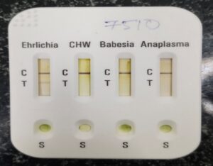
Figure 1. Tick Panel Combo SNAP Test showing a positive result for Ehrlichia.
Peripheral Blood Smear Examination
For a cytological examination, a blood smear was prepared from the blood collected for the tick-panel combo 4 SNAP test and allowed to air dry. The slide was stained with a Romanowsky-type stain and examined microscopically. Examination of the blood smear at low power (x100 objective) and high power (x40 objective), and scanning with an immersion oil lens (x100 objective), revealed intracytoplasmic basophilic inclusions characteristic of Ehrlichia morulae.
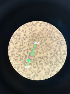
Figure 2. Cytological examination of peripheral blood smear showing ehrlichial morulae.
The presence of morula-like inclusions, together with the positive SNAP test and compatible clinical and hematological findings, provided further support for the diagnosis of Canine Ehrlichiosis. Although direct identification of morulae in a peripheral blood smear has limited sensitivity, their demonstration in an appropriate clinical setting is considered supportive evidence of infection (Harrus & Waner, 2011).
Treatment and Management
The dog received Oxytetracycline (OTC), Toxol, Lasix, and Ringer’s lactate as intravenous fluids for three days. The dog was then put on Doxycycline 10mg/kg b.wt (Doxypet®) for next 27 days, Pooch Liv-Guard®, and Pooch Nefro® syrup for a month as supportive therapy. Because of the renal and hepatobiliary involvement, the dog was put on a liver and kidney diet.
Table 4. Treatment protocol followed in the present case
| Medication | Form | Route | Purpose |
| OTC | Injection | I/V | Supportive therapy |
| Toxol | Injection | I/V | Supportive therapy |
| Lasix | Injection | I/V | Diuretic therapy |
| Ringer’s lactate | Infusion | I/V | Fluid therapy |
| Doxycycline (Doxypet®) | Tablet | Oral | Anti-Ehrlichial therapy |
| Pooch Liv-Guard® | Tablet | Oral | Hepatic supportive therapy |
| Pooch Nefro® | Syrup | Oral | Renal supportive therapy |
Discussion
The diagnosis of Canine Ehrlichiosis was made based on clinical findings, hematological and serum biochemical test results, ultrasonography, Tick-panel combo 4 snap test, and peripheral blood smear examination. Similar clinical signs, including anorexia, lethargy, weakness, and pale mucous membranes, have been reported in previous studies of Canine Ehrlichiosis (Neer et al., 2002). Anemia is a common finding in canine Ehrlichiosis and was observed in this case as evidenced by reduced hemoglobin levels, low RBC count, and reduced PCV. Anemia in Ehrlichiosis is attributed to bone marrow suppression, immune-mediated destruction of blood cells, and inflammatory processes (Mylonakis & Theodorou, 2017). Leukocytosis is also a common finding in canine Ehrlichiosis and was observed in this case.
However, thrombocytopenia, which is also commonly seen in canine Ehrlichiosis, was absent in this case since the platelet count was within the reference range. The serum biochemical test results showed hepatobiliary alterations, which were confirmed by ultrasonography. Similar to findings of (Mylonakis & Theodorou, 2017), marked increases in alkaline phosphatase and bilirubin levels were observed, suggesting hepatic dysfunction.
Renal involvement was also evident in this case, as demonstrated by markedly elevated urea, BUN, creatinine, and phosphorus levels, which were suggestive of severe azotemia. Similar to reports of (Ettinger et al., 2024; Verma et al., 2024), renal changes in canine Ehrlichiosis may be attributed to immune complex-mediated glomerulonephritis, inflammatory processes, and vasculitis. Finally, the detection of Ehrlichia morulae in this case provided direct evidence for the diagnosis of canine Ehrlichiosis. The presence of morulae in a peripheral blood smear is an important finding in the diagnosis of Canine Ehrlichiosis, although it is not always easy to demonstrate (Harrus & Waner, 2011). On the whole, hematological and serum biochemical tests and Tick-panel combo 4 snap test provided valuable information for the diagnosis of this case.
Conclusion
Canine Ehrlichiosis is a tick-borne disease that may cause serious clinical effects including anemia, renal failure, and hepatitis. The diagnosis of this disease depends on a combination of hematological and serum biochemical tests, ultrasonography, tick-panel combo 4 snap test, and microscopic examination of peripheral blood smear. The early diagnosis and treatment of this disease are critical to improve patient outcomes.
REFERENCES
Aziz, M. U., Hussain, S., Song, B., Ghauri, H. N., Zeb, J., & Sparagano, O. A. (2022). Ehrlichiosis in dogs: A comprehensive review about the pathogen and its vectors with emphasis on South and East Asian countries. Veterinary sciences, 10(1), 21.
Ettinger, S. J., Feldman, E. C., & Côté, E. (Eds.). (2024). Ettinger’s Textbook of Veterinary Internal Medicine-eBook: Ettinger’s Textbook of Veterinary Internal Medicine-eBook. Elsevier Health Sciences. Ettinger, S. J., Feldman, E. C., & Côté, E. (Eds.). (2024). Ettinger’s Textbook of Veterinary Internal Medicine-eBook: Ettinger’s Textbook of Veterinary Internal Medicine-eBook. Elsevier Health Sciences.
Groves, M. G., Dennis, G. L., Amyx, H. L., & Huxsoll, D. L. (1975). Transmission of Ehrlichia canis to dogs by ticks (Rhipicephalus sanguineus). American journal of veterinary research, 36(7), 937-940.
Harrus, S., Kass, P. H., Klement, E., & Waner, T. (1997). Canine monocytic ehrlichiosis: a retrospective study of 100 cases, and an epidemiological investigation of prognostic indicators for the disease. Veterinary Record, 141(14), 360-363.
Harrus, S., & Waner, T. (2011). Diagnosis of canine monocytotropic ehrlichiosis (Ehrlichia canis): an overview. The Veterinary Journal, 187(3), 292-296.
Mylonakis, M. E., & Theodorou, K. N. (2017). Canine monocytic ehrlichiosis: An update on diagnosis and treatment. Acta Veterinaria, 67(3), 299-317.
Neer, T. M., Breitschwerdt, E. B., Greene, R. T., & Lappin, M. R. (2002). Consensus statement on ehrlichial disease of small animals from the infectious disease study group of the ACVIM. Journal of Veterinary Internal Medicine, 16(3), 309-315.
Sykes, J. E. (2022). Greene’s Infectious Diseases of the Dog and Cat-E-Book. Elsevier Health Sciences.
Verma, S., Srivastava, M. K., Tiwari, J., Srivastava, A., Sachan, R., Bhatt, S., … & Singh, S. (2024). Study of Haematological and Biochemical Alterations in Clinical Cases of Canine Ehrlichiosis to Understand the Clinical Picture of the.
Waner, T., Harrus, S., Jongejan, F., Bark, H., Keysary, A., & Cornelissen, A. W. (2001). Significance of serological testing for ehrlichial diseases in dogs with special emphasis on the diagnosis of canine monocytic ehrlichiosis caused by Ehrlichia canis. Veterinary parasitology, 95(1), 1-15.