


Contagious pustular dermatitis: Etiopathology, diagnosis, clinical management and control
Das T1, Sahoo M1, Vidya Rani HB2, Chauhan A1 and Das NK3
1ICAR-NIFMD, Bhubaneswar
2PhD Scholar, ICAR-IVRI, Izatnagar
3BVO, Bisoi, Mayurbhanj, FARD, Government of Odisha
Corresponding author E mail id: tarenisahoo@gmail.com
Contagious pustular dermatitis (Orf) is a major obstacle and welfare problem ofsmall ruminants worldwide. It is an acute highly contagious debilitating economically important disease of sheep and goat. It can affect other animal species like camel, cattle, dogs and wild animals. It is a zoonotic disease having occupational hazard. Virus transmission takes place via direct contact with infected animals or indirect contact with infected dried scab present in the animal surroundings. There is also a possibility for saliva borne and milk borne transmission of Orf. Laboratory diagnosis is required to differentiate it from other similar or related condition or disease. There is no specific treatment for this disease. But treatment with broad spectrum parenteral antibiotics and other supportive / symptomatic treatment can make recovery faster. Ethnoveterinary preparation is also effective. Good managemental practices, strict hygiene, sanitation and biosecurity are required to prevent transmission of the disease.
Introduction
In Indian agricultural economy, small ruminants play a crucial role. Small ruminant production has emerged as an important segment in escalating and diversifying Indian agriculture. Small ruminants rearing is an integral part of rural living and about 70% of rural population belongs to landless and the marginal farmers. Small ruminants can adopt to harsh environment and act as life line during droughts, floods etc. They have short gestation period, higher feed conversion efficiency and are natural reproducible assets. Small ruminants rearing generates continuous source of employment and income for products like nutrition rich milk, meat, wool, hide, manure etc. and had positive impact on equity. The expanding population, urbanization and improved incomes have resulted in increase in consumption of animal products like meat and dairy and sustainable food production has important role in global food security. In India, small ruminant population was 223.2 million (148.9 million goats and 73.4 million sheep) in 2019 (20th-livestock-census). Total small ruminants’ population has increased as compared to previous livestock census 2012. However, there are many challenges or obstacles to small ruminant production including infectious diseases causing enormous economic losses. Contagious pustular dermatitis (Orf) is a major obstacle and welfare problem in small ruminants worldwide.
Etiology
Contagious pustular dermatitis is also called as sore mouth, scabby mouth, infectious labial dermatitis or contagious ovine ecthyma. It is an acute highly contagious debilitating economically important disease of small ruminants like sheep and goat. It is generally severe in goats than sheep. It can affect other animals like camel, cattle, dogs and wild animals. It is classified as notifiable disease by world organization of animal health. It is a zoonotic disease having occupational hazard posing greater risk to public health (Peralt et al., 2023). Animal handlers, meat handlers and veterinarians are at risk. It is caused by Orf virus (ORFV), genus Parapoxvirus, Sub-family Chordopoxvirinae, family Poxviridae (Gelaye et al., 2016). It is a large enveloped linear double stranded DNA virus 134kb-140kb in length. The genome consists of central core which encodes factors for virus replication and transcription and two inverted terminal repeats on both sides which encode factors playing important role in pathogenesis like induction of inflammation, immune evasion and host range (Kassa et al.,2021). The ORFV B2L gene encoding major envelope protein p37K is widely used for its taxonomic classification (Ghazaleh et al., 2023). Only 14 complete genome sequence of Orf virus is available (Sahu et al., 2022). The genomes OV-IA82 and OV-SA00 in America, NZ2 in New Zealand, D1701 in Germany, NA1/11 in China, IND/MP/17 from India have been completely sequenced (Zhang et al., 2016; Sahu et al., 2022).
The disease is characterized by proliferative lesions in mouth and muzzle and usually resolves in around 1-2 months. The disease is found worldwide. The disease has been reported across various Indian states like Uttarakhand, Assam (Bora et al., 2012), Uttar Pradesh (Kumar et al., 2014), Tripura (Venkatesan et al., 2018), Meghalaya, Rajasthan, Kashmir, Tamil Nadu (Nagarajan et al., 2019), Odisha (Sahu et al., 2019), Andaman and Nicobar Island (Sunder et al., 2019), Haryana etc. (Kumar et al., 2022). In Haryana, India in 2021, six outbreaks were reported in goats and only in one outbreak, sheep was affected (Kumar et al., 2022). The morbidity rate was 8.75% to 100% among goats which was consistently higher among young animals and the mortality rate was 2.5% to 60%. The morbidity rate in sheep was 0% to 8% (Kumar et al., 2022). The seropositivity rate was 76.62% among goats in Assam using indirect ELISA and highest antibody prevalence was reported among goats above 8 months old (Bora et al., 2016). The disease occurrence is more common during late summer, fall and winter. The disease is prevalent worldwide. De Oliveria et al., 2012 first reported severe Orf virus case in South America. In eastern region of Peninsular Malaysia, the reported overall seroprevalence was 22.8% (Bala et al., 2019). In Nigeria, 69.54 % farms were reported to have Orf during 2014-2016 with 25% morbidity and 15% mortality (Adedeji et al., 2022).
Transmission
The virus is very hardy and stable in the environment and can be recovered from dried scab after many years. Virus transmission takes place via direct contact with infected animals or indirect contact with infected dried scab present in the animal surroundings or indirect contact with contaminated feed, vehicles, fomites etc. (Chaudhary et al., 2022). House flies also mechanically transmit Para poxvirus (Shimuzi et al., 2022). Persons handling animals usually transmit the virus. Cat scratch can also cause ORFV infection (Frandsen et al., 2011). Nosocomial outbreak of ORFV in humans had also been reported in burn unit, Turkey having compromised skin integrity (Midilli et al., 2013). In a recent study, existence of saliva borne and milk borne transmission of infectious ORFV were described (Ma et al., 2022). Suckling of infected milk by newborn kids results in rapid outbreak of ORFV among them. Saliva contaminated mineral licks, feeder, waterer etc. can transmit ORFV from infected animals.
Viral virulence factors
The virus has developed certain evasive mechanism to evade active immune response, to grow and repeatedly infect the same host. Viral vascular endothelial growth factor plays important role in angiogenesis during skin infection (Savory et al., 2000). The viral VEGF induces fibroblast growth factor, keratinocyte growth factor, heparin binding epithelial growth factors which induces epidermal proliferation and rete ridges formation. The Orf virus with VEGF gene deletion produces no scab (Savory et al., 2000). The scab has important role in protecting virus from environment for 1 year after shedding and helps in infecting naïve animals. Chemokine binding protein (CBP) is a critical virulent factor for Orf virus (OV/ ORFV) expressed earlier stage of infection. It disturbs chemokine gradient in the infected tissue and inhibits recruitment of leucocytes to the infected site (Fleming et al., 2017). Another virulence factor is GIF (GM-CSF & IL-2 inhibitory factor) which is an intermediate-late viral gene of Orf virus has inhibitory effect on host inflammatory response and host immune response against virus infection. The Orf virus IL-10 like gene, also a virulence factor is transcribed early and interfere with type-I immune response (Fleming et al., 1997). OV interferon resistance gene protects virus from antiviral state of host by inhibiting PKR (Double stranded RNA dependent kinase) pathways (Haig et al., 1998). ORFV125gene induces apoptosis in antigen presenting cells through CD95/CD95L pathways and inhibits primary T- cell response (Yu et al., 2022).
Pathology
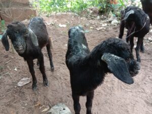
The infection is initiated through damaged skin, establishes infection in regenerated epidermal layer and progresses through different stages like erythema, papule, vesicle, pustule, scab etc. with infection confined to epidermis without any systemic spread (Savory et al., 2000). The incubation period is usually 3-7 days. The disease is self-limiting in adult animals usually resolves within 1-2 months. But in kids and lambs, it can be fatal (Ma et al., 2022). The skin lesions usually begin at oral commissures and later spread to muzzle and oral cavity (Lawan et al., 2020) and are very painful. The important clinical signs in the affected animals were pustular, proliferative, necrotic and scabby lesions around mouth, nostril and ears, anorexia and weakness. The oral lesions varying from small erythematous papule to large coalescing papules and ulcers were reported in the gingiva, tongue, hard palate, dental pad of young lambs (McElroy MC, Bassett HF, 2007). The lesions around mouth are associated with poor growth in young animals. Other clinical signs were peeling of skin, oozing of blood from hooves, nasal discharge, vesicles in oral cavity, glossitis, diarrhea, respiratory distress etc. (Kumar et al., 2022). In some cases, body parts like feet, udder and genitals like vulva, scrotum etc. were also affected leading to lameness and bacterial mastitis. Systemic spread of infection may result in severe gastroenteritis and severe bronchopneumonia. Secondary bacterial infection and maggot/ screwworms infestation always complicates the clinical outcome. Leukocytosis along with lymphocytosis and neutrophilia was reported in infected cases due to hosts response to inflammatory lesions initially and secondary bacterial infection later respectively.
The lesions have extensive vascular proliferation and dilation, extensive proliferation of epidermis with finger like projection rete ridges into the dermis with severe infiltration of inflammatory cells in the dermis (Savory et al., 2000). Hyperkeratosis, hydropic degeneration with presence of intracytoplasmic inclusion bodies and necrosis of keratinocytes, acanthosis, sub corneal pustules, intercellular oedema in the dermis with severe cellular infiltration were observed histopathologically (Devari et al., 2015). Severe persistent Orf was also reported in young Boer breed of goats with moderate to severe lymphadenopathy, suppurative arthritis, chronic fibrinous pneumonia, premature thymic involution, infestation of skin wounds with larvae (de la Concha-Bermejillo et al., 2003).
In humans, it is manifested by severe pain and development of pustules on hands and arms which can spread to other organs like perineal locations, genitals, face (Duchateau et al., 2014) and rarely on the scalp (Juang et al., 2023) and usually heal within 3-4 weeks in most people.
|
|
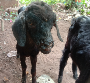 |
| Fig. 1: Kids affected with contagious ecthyma | |
Laboratory diagnosis
Serum neutralization test and counter immune electrophoresis are conventional method for Orf diagnosis (Sawhney et al., 1973). Polymerase chain reaction and semi nested PCR were developed for laboratory and differential diagnosis Orf viruses (Inoshima et al., 2000; Torfason and Gunadóttir, 2002). Sequencing and phylogenetic analysis of B2L gene encoding major envelope protein of ORFV helps to know genetic relatedness of the virus (Lojkic et al., 2002). Real time PCR assay based on TaqMan technology and a loop mediated isothermal amplification (LAMP) were developed for Orf virus detection (Gallina et al., 2006; Tsai et al., 2009; Bora et al., 2011). Virus can be isolated from scab suspension in primary or secondary lamb testes cell or lamb kidney cell line with production of typical cytopathic effect like cell rounding, ballooning, increased retractability and degeneration of cells (Venkatesan et al., 2011). For rapid detection of virus within 25 minutes, an ORFV-isothermal recombinase polymerase amplification (RPA)-lateral flow assay (LFA) with good correlation with q PCR was developed (Yang et al., 2016). A Dot ELISA was developed for successful detection OV in clinical samples (Nashiruddullah et al., 2022). Recombinant envelope protein based F1L indirect ELISA was developed with high sensitivity and specificity for detection of viral antibodies against OV (Yogisharadhya et al., 2018). Transmission electron microscopy was employed for ORFV detection and for describing scab lesion by immunogold electron microscopy and negative staining (Nashiruddullah et al., 2018).
Clinical management
There is no specific treatment for this disease. But treatment with broad spectrum parenteral antibiotics and other supportive / symptomatic treatment with anti-inflammatory, anti-histaminic, antipyretic, analgesic along with topical application of boric acid and desi curd paste on skin lesions or antiseptic like povidone iodine, boroglycerine or fly repellant ointment and honey on oral lesions can make recovery faster without any complication of secondary bacterial infection (Dar et al., 2015; Shivaraju et al., 2021). The lesions can be washed with 0.1% potassium permanganate solution before applying antiseptic and animal shed should be washed with 4% NaOH to prevent virus spread (Dalai et al., 2021). Aerosol herbal spray can be sprayed on the affected area. Weak animals can be provided with intravenous glucose saline and parenteral multivitamins like AD3E/ Belamyl (Chaudhary et al., 2022). In case of diarrhea, anti-diarrheal can be provided.
Ethnoveterinary preparation made from turmeric powder and aloe vera gel (1:1) was also found effective in Orf treatment (Brahma et al., 2020). Homemade preparation of turmeric and ghee is also effective (Chaudhary et al., 2022).
Control and prevention
Good managemental practices are very crucial in prevention and control of this diseases. The animal should be provided with nutritious diet and sufficient drinking water. The animal houses should be well ventilated, well-constructed and should be routinely cleaned and disinfected to prevent overcrowding and disease transmission. Orf is difficult to control after it enters into farm. So, quarantine of new animals is necessary before their introduction to farm. Infected animals in a farm should be isolated. The farm equipment, fomites and surroundings should be properly disinfected during outbreak. The infected animals should be handled lastly after handling healthy animals. the younger animals should be tube fed. The animals should not be fed with harsh vegetation as it causes skin injury. During the outbreak, movement of living and non-living things in and out of farm should be avoided. Awareness on these enzootic diseases needs to be created among public. The animal handlers should use protective measures like gloves, shoes cover, masks etc. while handling animals. All the infected materials should be properly incinerated. Strict hygiene, sanitation and biosecurity are required to prevent transmission of the disease. The milk and saliva from infected and asymptomatic dairy animals have been highlighted as potential novel source of ORFV transmission. Pasteurization and heat treatment of milk can inactivate live virus and prevent further transmission. Farm workers should wear protective measures while milking and handling raw milk (Ma et al., 2022). Arthropod control measures in farms are necessary to prevent mechanical transmission of ORFV by house flies. The live virus vaccine is available to prevent ORF. Commercial vaccines are available for ORF prevention. As this contains live virus, vaccination should be done in problematic herd during outbreak only. The vaccinated animals should be isolated from unvaccinated animals until scab falls down. Vaccinators should take precautions and wear gloves while handling vaccine.
Reference
Abu Ghazaleh R, Al-Sawalhe M, Abu Odeh I, El Ibrahim J, Al-Turman B, Makhamreh J. (2023). Host range, severity and trans boundary transmission of Orf virus (ORFV). Infect Genet Evol. 112:105448.
Adedeji AJ, Adole JA, Asala OO, Gamawa AA, Maurice NA, Jambol A, Bolajoko MB, Chima NC, Ifende VI, Wungak YS, Woma TY, Luka PD. (2022). A survey of contagious ecthyma and molecular characterization of Orf virus in sheep and goats in Nigeria (2014-2016). Open Vet J.12(4):551-561
Bala JA, Balakrishnan KN, Abdullah AA, Adamu L, Noorzahari MSB, May LK, Mangga HK, Ghazali MT, Mohamed RB, Haron AW, Noordin MM, Lila MAM. (2019). An association of Orf virus infection among sheep and goats with herd health programme in Terengganu state, eastern region of the peninsular Malaysia. BMC Vet Res. 18;15(1):250
Bora DP, Barman NN, Das SK, Bhanuprakash V, Yogisharadhya R, Venkatesan G, Kumar A, Rajbongshi G, Khatoon E, Chakraborty A, Bujarbaruah KM. (2012). Identification and phylogenetic analysis of orf viruses isolated from outbreaks in goats of Assam, a northeastern state of India. Virus Genes. 45:98-104.
Bora DP, Venkatesan G, Bhanuprakash V, Balamurugan V, Prabhu M, Sankar MS, Yogisharadhya R. (2011). TaqMan real-time PCR assay based on DNA polymerase gene for rapid detection of Orf infection. Journal of virological methods. 178(1-2):249-52.
Bora M, Bora DP, Barman NN, Borah B, Das S. (2016). Seroprevalence of contagious ecthyma in goats of Assam: An analysis by indirect enzyme-linked immunosorbent assay. Vet World. 9(9):1028-1033.
Brahma J, Saharia J, Sarma M, Boro P. (2020). Successful treatment of contagious ecthyma (ORF) in Assam hill goats by using turmeric powder and aloe vera gel preparation. J Entomol Zool Stud. 8:1454-6.
Dalal J, Sangwan A, Kumar A, Yadav R. (2021). Molecular diagnosis and therapeutic management of contagious ecthyma in beetle goat. Haryana Vet. 60(2), 298-300
Dar KH, Tufani NA, Dar SH, Hafiz A, Naikoo MD. (2015). Comparative therapeutic management of contagious ecthyma in small ruminants. Intas Polivet. 16(2):431-5..
Davari SA, Sayyari M, Mohammadi A. (2015). Histopathological study and F1L gene sequence analysis of contagious ecthyma in small ruminants of Shiraz suburb, Iran. Trop Biomed. 32(2):335-43.
de la Concha-Bermejillo A, Guo J, Zhang Z, Waldron D. (2003). Severe persistent orf in young goats. J Vet Diagn Invest. 15(5):423-31.
de Oliveira CH, Assis FL, Neto JD, Oliveira CM, Lopes CT, Bomjardim Hdos A, Vinhote WM, Silva AG, Abrahão JS, Kroon EG. (2012). Multifocal cutaneous ORF virus infection in goats in the Amazon region, Brazil. Vector Borne Zoonotic Dis. 12(4):336-40.
Duchateau NC, Aerts O, Lambert J. (2014). Autoinoculation with Orf virus (ecthyma contagiosum). International Journal of dermatology. 53(1):e60-2.
Fleming SB, McCaughan C, Lateef Z, Dunn A, Wise LM, Real NC, Mercer AA. (2017). Deletion of the Chemokine Binding Protein Gene from the Parapoxvirus Orf Virus Reduces Virulence and Pathogenesis in Sheep. Front Microbiol. 24;8:46.
Fleming SB, McCaughan CA, Andrews AE, Nash AD, Mercer AA. (1997). A homolog of interleukin-10 is encoded by the poxvirus orf virus. J Virol. 71(6):4857-61.
Frandsen J, Enslow M, Bowen AR. (2011). Orf parapoxvirus infection from a cat scratch. Dermatol Online J.17(4):9.
Gallina L, Dal Pozzo F, Mc Innes CJ, Cardeti G, Guercio A, Battilani M, Ciulli S, Scagliarini A. (2006). A real time PCR assay for the detection and quantification of orf virus. J Virol Methods. 134(1-2):140-5.
Gelaye, E., Achenbach, J.E., Jenberie, S. et al. (2016). Molecular characterization of orf virus from sheep and goats in Ethiopia, 2008–2013. Virol J 13, 34
Haig DM, McInnes CJ, Thomson J, Wood A, Bunyan K, Mercer A. (1998). The orf virus OV20.0L gene product is involved in interferon resistance and inhibits an interferon-inducible, double-stranded RNA-dependent kinase. Immunology. 93(3):335-40.
Inoshima Y, Morooka A, Sentsui H. (2000). Detection and diagnosis of parapoxvirus by the polymerase chain reaction. Journal of virological methods. 84(2):201-8.
Juang SJ, Win KT, Chen YL, Chen HW, Cheng PS. (2023). Orf Infection on the Scalp of a Taiwanese Woman: A Case Report and Literature Review. Life (Basel). 28;13(2):358.
Kassa T. (2021). A Review on Human Orf: A Neglected Viral Zoonosis. Res Rep Trop Med. 12:153-172.
Kumar N, Wadhwa A, Chaubey KK, Singh SV, Gupta S, Sharma S, Sharma DK, Singh MK, Mishra AK. (2014). Isolation and phylogenetic analysis of an orf virus from sheep in Makhdoom, India. Virus Genes. 48:312-9.
Kumar R, Moudgil P, Grakh K, Jindal N, Sharma M, Gupta R. (2022). Epidemiology, clinical features, and molecular detection of orf virus in Haryana (India) and its adjoining areas. Trop Anim Health Prod. 18;54(5):268.
Lojkic I, Cac Z, Beck A, Bedekovic T, Cvetnic Z, Sostaric B. (2010). Phylogenetic analysis of Croatian orf viruses isolated from sheep and goats. Virol J. 7:314.
Ma W, Pang M, Lei X, Wang Z, Feng H, Li S, Chen D. (2022). Orf Virus Detection in the Saliva and Milk of Dairy Goats. Front Microbiol13:837808.
McElroy MC, Bassett HF. (2007). The development of oral lesions in lambs naturally infected with orf virus. Vet J. 174(3):663-4.
Midilli K, Erkiliç A, Kuşkucu M, Analay H, Erkiliç S, Benzonana N, Yildirim MS, Mülayim K, Acar H, Ergonul O. (2013). Nosocomial outbreak of disseminated orf infection in a burn unit, Gaziantep, Turkey, October to December 2012. Euro Surveill. 18(11):20425.
Nagarajan G, Pourouchottamane R, Reddy GM, Yogisharadhya R, Sumana K, Rajapandi S, Murali G, Thirumaran SM, Mallick PK, Rajendiran AS. (2019). Molecular characterization of Orf virus isolates from Kodai hills, Tamil Nadu, India. Veterinary World. 12(7):1022.
Nashiruddullah N, Pathak DC, Barman NN, Ahmed JA, Begum SS, Roychoudhury P. (2022). Natural infection of goats with orf (Contagious ecthyma) and its diagnosis. Indian Journal of Animal Research. 56(2):192-200.
Nashiruddullah N, Pathak DC, Barman NN, Ahmed JA, Borah P, Begum SS, Islam S. In vitro and in vivo assessment of orf virus (ORFV) by electron microscopy. (2018). Veterinarski arhiv. 88(6):847-61.
Peralta A, Flores-Olivares C, Verna A, González-Altamiranda E, Odriozola E, Madariaga C, Odeón A, König GA, Cantón G. (2023). Identification and molecular characterization of Orf virus infection in occupationally exposed women in South America. Rev Argent Microbiol. 55(2):129-132.
Sahu BP, Majee P, Sahoo A, Nayak D. (2019). Molecular characterization, comparative and evolutionary analysis of the recent Orf outbreaks among goats in the Eastern part of India (Odisha). Agri Gene. 12:100088.
Sahu BP, Majee P, Singh RR, Sahoo N, Nayak D. (2022). Recombination drives the emergence of orf virus diversity: evidence from the first complete genome sequence of an Indian orf virus isolate and comparative genomic analysis. Arch Virol. 167(7):1571-1576
Savory LJ, Stacker SA, Fleming SB, Niven BE, Mercer AA. (2000). Viral vascular endothelial growth factor plays a critical role in orf virus infection. J Virol. 74(22)
Sawhney AN, Dubey SC, Malik BS. (1973). Diagnosis of contagious pustular dermatitis in sheep and goats by agar-gel precipitation test. Indian veterinary journal.
Shimizu K, Takase H, Okada A, Inoshima Y. (2022). Possibility of mechanical transmission of parapoxvirus by houseflies (Musca domestica) on cattle and sheep farms. J Vet Med Sci. 84(9):1313-1319.
Shivaraju S, Mohan D, Kalaiselavn E, Maiti S, Prakash G. (2021). Successful clinical management of contagious ecthyma (Orf) in goat: A case report. Int J Vet Sci Anim Husbandry. 6(1):41-2.
Sunder J, Sujatha T, De AK, Bhattacharya D, Bhowmick S, Perumal P, Kundu A. First report of contagious ecthyma (orf) outbreak in goats of Andaman and Nicobar Islands.
Torfason EG, Gunadóttir S. (2002). Polymerase chain reaction for laboratory diagnosis of orf virus infections. J Clin Virol. 24(1-2):79-84.
Tsai SM, Chan KW, Hsu WL, Chang TJ, Wong ML, Wang CY. (2009). Development of a loop-mediated isothermal amplification for rapid detection of orf virus. J Virol Methods. 157(2):200-4.
Venkatesan G, Balamurugan V, Bora DP, Yogisharadhya R, Prabhu M, Bhanuprakash V. (2011). Sequence and phylogenetic analyses of an Indian isolate of orf virus from sheep. Vet Ital. 47(3):323-32.
Venkatesan G, De A, Arya S, Kumar A, Muthuchelvan D, Debnath BC, Dutta TK, Hemadri D, Pandey AB. (2018). Molecular evidence and phylogenetic analysis of orf virus isolates from outbreaks in Tripura state of North-East India. Virusdisease. 29:216-20.
Yang Y, Qin X, Wang G, Jin J, Shang Y, Zhang Z. (2016). Development of an isothermoal amplification-based assay for rapid visual detection of an Orf virus. Virol J. 13:46.
Yogisharadhya R, Kumar A, Bhanuprakash V, Shivachandra SB. (2018). Evaluation of a recombinant major envelope protein (F1L) based indirect- ELISA for sero-diagnosis of orf in sheep and goats. J Virol Methods. 261:112-120
Yu Y, Lian Z, Cui Y. (2022). The OH system: A panorama view of the PPV-host interaction. Infect Genet Evol. 8:105220.
Zhang K, Xiao Y, Yu M, Liu J, Wang Q, Tao P, Liu S, Ning Z. (2016). Phylogenetic analysis of three orf virus strains isolated from different districts in Shandong Province, East China. J Vet Med Sci. 77(12):1639-45.
A Mini Review on Diagnosis and Treatment of Lumpy Skin Disease (LSD) Menace in Cattle in India