

Diagnosis of Microfilaria Infection in Dog with Geimsa stain Technique -A Case Report
Dr.Bimal Bey
Parasitology Section
Division of Animal Health, ICAR Research Complex for NEH Region, Umiam,
Meghalaya. -793103
Case Report:
In this report blood sample from a 6-year-old Labrador dog was collected which was reported by owner from Barapani, Umroi road to the ICAR Research Complex Umiam, Meghalaya and had the previous history of anorexia, weakness, coughing and dyspnoea. During the physical examination the dog had shown debility, Pale mucous membrane and high rise of body temperature (39 degrees Celsius), laboured breathing, weakness but had found normal respiratory and heart rate.
With the help of disposable syringe, a blood sample (2 ml) from cephalic vein of the infected dog was collected in an anticoagulant containing (EDTA) vial. The blood sample was examined with Geimsa stain technique with staining.
In the Giemsa stain technique, a thin blood smear is made in a slide and staining with Giemsa stain and slide was kept it for air dry. After that, it was examined on low power (40X) microscope and found the present of microfilaria an elongated snake (fig) like structure.
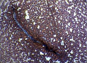
Figure: Shown the microfilaria worm under the Geimsa stain technique.
Treatment:
Effective treatment of doxycycline at rate of 10 mg/kg orally twice daily for 30 days and combined treatment with ivermectin 6 mg/kg orally for weekly interval recovered eventually in 30 days after beginning of the treatment.
Result and Discussion:
The used of effective method of blood smear with Geimsa stain diagnosed the positive result of microfilaria infection.
In the present case report study, revealed that the vector borne disease such as microfilarisis are endemic in the sub-tropical region including north east India like Meghalaya, Mizoram and Assam. These states are vulnerable to climate change characterized by increased rainfall and environmental temperature resulting likelihood increase of vector borne diseases including human malaria. Dirofilaria immitis (Nematoda: Filariidae) causes heartworm disease, which is widespread throughout tropical and temperate regions of the world including Europe stated by Mario Santoro et al. (2019).
Transmitted from host-to-host via mosquito bites and the prevalence of the disease is influenced by climate and topography, but most importantly, by the presence of mosquito vectors. Nguyenet et al.(2016).
The owner from Barapani, Umroi road was reported a Labrador dog to the ICAR Research Complex Umiam, Meghalaya and the owner also informed the bites of mosquitoes and the dog was shown the symptoms of micro filariasis and in this report the result found was also similar with Sonjoy Kumar Borthakur et al (2014). He was also found the main clinical symptoms in dirofilariosis include persistent cough, difficult breathing, and poor exercise tolerance followed by ascites, anorexia, and weight loss.
In this report, the history of mosquito’s bite which was similar with finding by A.L. Vieira et al, 2014.Where they had reported the different species of culicid mosquitoes belonging to the genera Culex, Aedes, and Anopheles, among others, have been implicated in the transmission of this parasite, allowing for its intermediate stage to complete its life cycle.
In the present case report microfilariasis was diagnosed with the helped of Giemsa staining technique as shown in the figure, microfilaria under the low power (40X) microscope. This was similar with Ibeh Nnanna Isaiah et al. (2021). Were identified D. Immitis with the helped of Giemsa stained blood smears technique.
At presence case report the dog was advised to treat with doxycycline at rate of 10 mg/kg orally twice daily for 30 days. Blood samples were collected every 30 days during 6 months of treatment and combined treatment of doxycyclin with ivermectin 6 mg/kg orally for weekly was given. This treatment result was also similar with Amarjit et al. (2017), where the combine treatment of Microfilaria infection in dog with ivermectic injection of at the rate of 0.3mg/kg body weight subcutaneously weekly for four occasions with doxycycline the rate of 10mg/body weight orally for one month was effective.
Summary:
The used of effective method of blood smear with Geimsa stain diagnosed the positive result of microfilaria infection.
Acknowledgement:
The author is thankful to the Scientist Dr. R. Laha and Dr. M. Das, Parasitology Section, Division of Animal Health, ICAR Research Complex for providing all the necessary facilities.
References:
Amarjit et al.(2017). Doual infection of Microfilaria and Bebesia in a Labrador dog in Meghalaya.Indian journal of canine practice. (9) 114-116.
A.L. Vieira et al. (2014). Prevalence of canine heartworm (Dirofilariaimmitis) diseasein dogs of Central Portugal.Parasite 21 (5):1-7.
Mario Santoro et al. (2019).Heartworm Disease (Dirofilaria immitis) in Two Roaming Dogs From the Urban Area of Castel Volturno, Southern Italy. Front Vet Sci. 2019; 6: 270.
Nguyen et al. (2016). Mosquito-borne heartworm Dirofilariaimmitis in dogs from Australia. Parasites & Vectors, 9:535-546.
Sonjoy Kumar Borthakur et al. (2014). Prevalence and Molecular Epidemiological Data on Dirofilariaimmitisin Dogs from Northeastern States of India.The ScientificWorldJournal.2015:1-7.
Ibeh Nnanna Isaiah et al. (2021). Evaluation of an improved Giemsa staining technique for blood borne parasite in thick and thin blood film from dogs. International Journal of Biology and Pharmacy Research Archive, 2021, 01(01), 001–005.