



Malignant Catarrhal Fever
Dr. Apurv Pandey, Dr. Maneesh Jatav, Dr. Yamini Verma, Dr. Amita Dubey and Dr. Madhu Swamy
Department of Veterinary Pathology,
College of Veterinary Science and Animal Husbandry,
Nanaji Deshmukh Veterinary Science University (NDVSU),
Jabalpur – 482001, Madhya Pradesh (M.P.), India
Corresponding Author: Dr. Apurv Pandey apupandey199@gmail.com
Introduction: Malignant catarrhal fever (MCF) is an infectious viral disease of ruminants and is usually fatal. Earlier, MCF was considered rare and sporadic in India, with only a few reported cases. However, in recent years, it has emerged as an important and increasing disease problem in the country. The disease is caused by a gamma herpesvirus, most commonly Ovine herpesvirus-2 (OvHV-2). Domestic sheep act as asymptomatic carriers of the virus, while infection in susceptible species results in severe clinical disease known as sheep-associated MCF (SA-MCF). Species commonly affected include cattle, deer, bison, water buffalo, and pigs. SA-MCF has been reported worldwide and can occur throughout the year. In India, mixed livestock farming practices, where cattle are reared alongside sheep and goats, are common. Such systems increase the risk of close contact between carrier animals and susceptible species during housing or grazing, thereby facilitating transmission of the virus.
Transmission: OvHV-2 is transmitted by contact or aerosol, from carrier lambs under 1-year old. The disease can also be transmitted by goats which also are the asymptomatic carriers of the disease. Incubation periods after experimental inoculation of cattle are 2-12 weeks. The clinical disease occurs when the susceptible host are in contact with asymptomatic sheep/goat either directly or indirectly. The respiratory tract is a common route for both entry and shedding of the virus. Infectious OvHV-2 is present in ovine nasal secretions, adult sheep can be infected by OvHV-2 via horizontal transmission through natural contact and it may intermittently shed large quantities of virus from nasal secretions which when transmitted to the susceptible cattle causes a fatal disease.
Pathogenesis: Sheep-associated malignant catarrhal fever (SA-MCF) in cattle is caused by a gamma herpesvirus that remains asymptomatic in its natural host (sheep) but induces a severe, frequently fatal disease in cattle.
- Transmission and Viral Entry: Cattle acquire OvHV-2 primarily through inhalation of virus-laden aerosols originating from nasal and ocular secretions of infected sheep, particularly lambs. Following exposure, the virus gains entry through the upper respiratory tract mucosa and is transported systemically via infected leukocytes.
- Cellular Tropism and Establishment of Latency: In cattle, OvHV-2 does not undergo productive replication. Instead, the virus establishes latent infection predominantly within CD8⁺ cytotoxic T lymphocytes. Viral genome persists episomally, with minimal viral gene expression, evading effective immune clearance.
- Immune Dysregulation and Lymphoproliferation: The central event in SA-MCF pathogenesis is aberrant activation and clonal expansion of OvHV-2-infected CD8⁺ T lymphocytes. These cells exhibit uncontrolled proliferation and heightened cytotoxic activity, leading to widespread tissue infiltration. Disease manifestations result primarily from immune-mediated injury, not direct viral destruction of host cells.
- Vasculitis as the Hallmark Lesion: Activated lymphocytes preferentially accumulate around small- and medium-sized blood vessels, causing severe lymphocytic vasculitis. Endothelial injury, fibrinoid necrosis, and perivascular inflammation compromise vascular integrity, leading to ischemia, hemorrhage, and tissue necrosis. This systemic vasculitis is the pathological hallmark of OvHV-2 infection in cattle.
- Vascular and lymphoid lesions result in widespread organ damage such as:-
- Oral and nasal mucosa show erosions, ulcers, and necrosis.
- Eyes show keratitis and corneal opacity (“blue eye”)
- Lymphoid tissues show marked lymphadenopathy due to lymphoid hyperplasia
- Central nervous system shows vasculitis and perivascular cuffing, leading to neurological signs
Clinical symptoms: The disease MCF occurs in different forms;
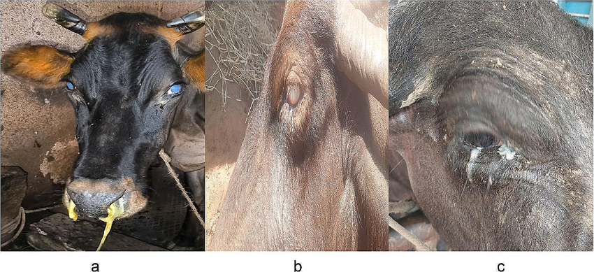
Head and eye form: – Most common and classical form of sheep-associated MCF.
- High, persistent fever (41-41.5 °C), severe depression, anorexia, and agalactia.
- Ocular lesions include bilateral keratoconjunctivitis with progressive corneal opacity (“blue eye”) (Fig. 02), lacrimation, blepharospasm, and congestion of scleral vessels, often leading to blindness.
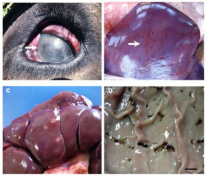
Figure 1: Corneal opacity
- Nasal and oral lesions show profuse mucopurulent nasal discharge, nasal obstruction causing dyspnea, erosions and necrosis of oral and nasal mucosa, gums, hard palate, tongue, and lips, painful mouth with excessive salivation and difficulty opening jaws.
Figure 2: Mucopurulent nasal discharge
- Lymphadenopathy is marked enlargement of superficial lymph nodes.
- Neurological signs include incoordination, weakness, muscle tremors, nystagmus and terminal stages show head pressing, paralysis, and convulsions.
- Disease shows rapid progression with death usually within 7-10 days.
- Peracute Form:- This form is rapidly progressive and often fatal, especially in young calves.
- Disease duration is of 1–3 days
- Sudden onset of very high fever
- Severe dyspnea
- Acute gastroenteritis
- Minimal development of classical mucosal or ocular lesions
- Death may occur before typical signs fully develop
- Alimentary Form:- In this form, gastrointestinal involvement predominates.
- Profuse watery or hemorrhagic diarrhoea
- Dysentery
- Rapid dehydration and weakness
- Oral and nasal erosions may be present but are less severe
- Eye lesions are minimal or absent
- Fever and lymphadenopathy are usually present
- Mild Form:- This form is rare and is observed mainly in experimentally infected animals, though occasional field cases occur.
- Transient fever
- Mild depression
- Mild erosions of oral and nasal mucosa
- Minimal ocular involvement
- Animals may survive the acute phase
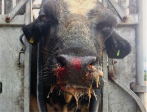
Figure 3: Mucopurulent and haemorrhagic nasal discharge with erosion to the muzzle and some periorbital swelling.
Pathology
- Gross Lesions
- Erosive and ulcerative lesions in oral, nasal, and gastrointestinal mucosa
- Enlarged lymph nodes
- Petechial hemorrhages in organs
- Histopathology
- Lymphocytic vasculitis
- Perivascular cuffing
- Necrosis of epithelial tissues
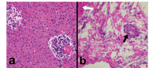
Figure 4: A) Liver. Severe vasculitis in the central veins. H&E. ×400; B) Heart. Epicardial vasculitis (black arrow). H&E. ×400
Diagnosis:
- Clinical Diagnosis: Based on characteristic signs,
- Pathological Diagnosis: Based on gross and histopathological lesions,
- Molecular diagnosis:
- Polymerase Chain Reaction Test has become an important tool in MCF molecular diagnostics.
- Recently real time PCR for OvHV-2 DNA has also been developed.
- A multiplex PCR has also been developed which can be used to diagnose the disease in different wild and domestic ruminants in one test only.
- Despite numerous efforts, the virus has not been isolated worldwide and therefore the demonstration of Genome by PCR is considered the gold standard for the diagnosis of the disease.
Differential diagnosis:
- Vesicular Stomatitis
- Infectious Bovine Rhinotracheitis (IBR)
- Bovine Viral Diarrhea / Mucosal Disease
Control and prevention: There is no treatment for the disease, symptomatic treatment rarely helps. There are no vaccines available till date. Since the main source of infection in these farms is the lambs, 9-12 months of age which shed virus in high quantities certain points to keep in mind for control of the disease are enlisted below:
- Segregation of the susceptible host from the reservoir hosts
- If cattle and sheep are reared under natural flock conditions, the grazing areas should be separate.
- It has been suggested that grazing sheep in the morning hours and cattle in the evening hours on the same land patch may also help as the virus may be destroyed in the day’s sun as it is very heat labile.
- A program to produce sheep free of infection should be initiated in which; lambs are removed from contact with infected sheep prior to 2 months of age.
- Zoological parks should only introduce sero-negative animals only and follow strict quarantine restrictions of newly acquired animals.
- Systematic surveillance program must be implemented with collaboration of national institutes like NISHAD and ICAR- NIVEDI.