


Mastitis: Prevention and Control
Sweety, Department of Physiology & Biochemistry, LUVAS, Hisar.
Pradeep Kumar, Department of Veterinary Medicine, LUVAS, Hisar.
https://www.pashudhanpraharee.com/prevention-of-mastitis-in-dairy-farm-by-good-management-practices/
Mastitis is a multifactoral disease, closely related to the production system and environment where cows are kept in. It is an inflammatory condition of the udder in which there are changes in the milk colour and consistency. Milk yield reduces abruptly which results in heavy economic losses. It is one of the most devastating diseases on dairy farms. High yielding dairy cows are more commonly affected than low yielders. Exotic and cross bred cows are more prone to mastitis than the Indian zebu cows. Heifers can also be affected by udder infections, even prior to calving.
Approximately 60% of all heifers have an intramammary infection at calving. Some 16% of these heifers will suffer from clinical mastitis during their first lactation and 30% of these mastitis cases will occur within 14 days after calving. This results in a reduced milk yield in the first lactation, causing severe economic losses.
- Causes
- A large number of species of microorganisms have been implicated as causes of mastitis. They are bacteria, fungus, Mycoplasma and virus. It usually occurs as an immune response to bacterial invasion of the teat canal by variety of bacterial sources present on the farm (commonly through bedding or contaminated teat dips), and can also occur as a result of chemical, mechanical, or thermal injury to the cow’s udder.
- The most important bacterial organisms causing mastitis are Staphylococcus aureus; Str. agalactiae; Str. zooepidemicus; faecalis; Str. pyogenes; Klebsiella spp; Mycobacterium bovis; E.coli.
- The fungal organisms responsible for mastitis are Trichosporon spp; Aspergillus fumigatus; A. midulus; Candida spp.
- Mode of Transmission
- Environmental: The cutaneous surface of the cow may have many organisms as resident population and from where the organisms may invade teat canal and infection reaches the mammary gland. The normal inhabitant of udder and environment like Agalactiae, Stap. aureus and E.coli under favourable conditions multiply and invade the tissues produce much damaging effect.
- Contamination during handling: The contamination of milker’s hands, clothes and machine cup by milk from the affected quarter may lead to the spread of the disease to other non-infected teats of cow.
- Flies and other insects may also spread the infection from one place to the other. Spread of infection is possible through bedding ground by discharges of affected gland.
- Symptoms
Clinical symptoms
- Swollen udder with hot, reddened, painful and hard teats.
- Animal will not allow touching the udder and will kick while touching it.
- Milk mixed with blood which give reddish tinge.
- Milk mixed with yellow or brown fluid with flakes or clots and foul smelling.
- Reduced milk yield.

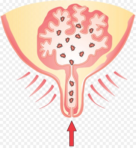
Swollen udder |
- Managemental Methods
- Concrete floor should be avoided and animal should be provided with soft bedding.
- Bedding should be of straw, saw dust or sand.
- Infusion should be used in each cow during dry period.
- Animal sheds should be kept clean.
- Udder and hands of the milker should be washed with antiseptic lotion (4% Potassium permanganate solution) before and after milking.
- Proper cleaning and disinfection of milking machine and the teat cup, vessels should be done after each milking.
- The healthy non-infected cows should be milked first and known infected cows should be milked at last.
- Newly introduced cow should be milked separately and should be screened through California Mastitis Test (CMT).
- Immediately after milking don’t allow the animal to lie-down by engaging with fodder.
- The complete milking should be done at every time and milk should not be stored in teats.
- The udder and teats should be protected from any injuries.
- Hygienic measures at milking time, udder preparation before milking, post milking teat disinfections have been recommended as preventive measures.
- Control of fly population should be attempted, for these insecticides fly repellent sprays are to be made in the house and surroundings.
- The frequently affected animals should be removed from the herd.
- Prevention and control
- Mastitis in heifers can be prevented. In the first place by managerial measures by eliminating the sources of infection.
- Reduce stress on the animals by maintaining proper nutrition, ventilation and housing.
- Maintain teat hygiene which includes good housing management, effective teat preparation and disinfection for good milk hygiene, teat health and disease control.
- Reduce the amount of bacteria in the environment (clean housing and bedding).
- Optimise insect control.
- Remove sucklers from groups of young stock.
- In addition, changes that reduce or eliminate risk factors associated with mastitis should be considered.
- Prompt identification and treatment of clinical mastitis cases should be done including the use of the most appropriate treatment for the symptoms.
- Dry cow management and therapy should be done where cows are dried off abruptly and teats are cleaned before dry cow antibiotics are administered, including the use of teat-end sealants.
- Regular testing and maintenance of the milking machine should be done with regular, recommended teat cup liner replacement and milking machine servicing and attention paid to items which must be checked on a daily, weekly or monthly basis.
- Good record keeping of all aspects of mastitis treatment, dry cow therapy, milking machine servicing, Somatic Cell Count results, and clinical mastitis cases.
Diagnosis and treatment
- Herd screening should be done on routine basis to detect subclinical, clinical mastitis. For this purpose California Mastitis Test (CMT) can be used and then somatic cell count and bacterial culture.
- Other tests like ELISA, bacterial culture and multiplex PCR can be used.
Conclusion
- Mastitis is caused by a variety of bacteria and many of these cases have a high rate of spontaneous cure.
- Many subclinical pathogens are responsive to intramammary treatments using commercially available antibiotic products but there are important cow & herd factors that will influence the cost effectiveness of treatment.
- The decision to treat subclinical mastitis is dependent upon the type of pathogens that are prevalent and diagnostic efforts (milk culturing) must be undertaken before developing a treatment protocol.
- Early detection of mastitis and its careful management is essential for the well-being of a dairy herd.