SURGICAL MANAGEMENT OF AURICULAR TUMOUR IN A DOE
- Vigneswari1*, P. Sankar2, A. Kumerasan2, S. Kathirvel2, K. Jayakumar2 and
- Vigneshwaran2
1Assistant Professor,
Department of Veterinary Surgery and Radiology
Rajiv Gandhi Institute of Veterinary Education and Research (RIVER),
Kurumbapet, Puducherry, India.
2 Department of Veterinary Surgery and Radiology
Veterinary College and Research Institute, Namakkal – 637 002 (Tamil Nadu)
Tamil Nadu Veterinary and Animal Sciences University (TANUVAS), India.
*Corresponding author
E mail ID: vigneswari.n0@gmail.com
Abstract
A four years old pleuriparous non-descript Doe was presented to the Veterinary Clinical Complex, Veterinary College and Research Institute, Namakkal with the history of unusual swelling in the base of left ear for the past 2 months and the goat was experiencing difficulty in moving its ear due to the weight of the tumour. Clinical examination of the swelling revealed hard in consistency and non-fluctuating. All physiological and haemato-biochemical parameters were within normal range. The mass was tentatively diagnosed as tumour. The tumour mass was surgically excised and sample was sent for histopathology. Surgical wound was closed routinely. Antibiotic, analgesics, tetanus toxoid were administered postoperatively and daily cutaneous wound dressing was performed. On 10th post-operative day sutures were removed and animal made an uneventful recovery.
Keywords: Aural fibroma, Doe.
Introduction
Fibromas are benign tumour that composed of fibrous or connective tissue. They can grow in all organs, arising from mesenchyme tissue and fibroma in pinna is uncommon. Neoplasia is occasionally diagnosed in goats. A survey of 800000 slaughtered goats revealed only 70 neoplasm’s (Bradly and Magaki, 1963). This paper describes the aural fibroma and its surgical excision in a non-descript doe.
Materials and methods
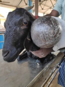
A four years old pleuriparous non-descript Doe with the history of unusual swelling in the base of left ear for the past 2 months. The goat was experiencing difficulty in moving its ear due to the weight of the tumour (Fig 1). The goat was sedated with Diazepam @ 0.5mg per kg body weight intravenously. Surgical site was prepared aseptically. Local infiltration anaesthesia was given using 2% lignocaine hydrochloride around the mass. An elliptical skin incision made around the base of the ear including tumour mass and separated (Fig 2). The tumour mass was surgically excised and sample was sent for histopathology and the case was confirmed as fibroma showing collagen rich fusiform to spindle shaped hyperchromatic fibroblasts arranged as interlacing bundles and whorls (Fig 3). The skin incision was closed excluding the cartilage using simple interrupted suture pattern. Postoperatively antibiotics, analgesics and wound care were given. On 10th postoperative day sutures were removed and animal made an uneventful recovery.
Results and discussion
Aural fibroma is a rare condition in goats. The excised mass was black in colour and hard in consistency. The tumour originated from cartilage of pinna. The weight of tumour was 380 gm. Histopathology revealed fibroblasts and their nuclei were spindle-shaped and arranged together with collagen fibers in a concentric manner forming whorls around the blood vessel. The goat recovered completely and no reoccurrence reported at present. Susan et al (2016) reported fibro epithelial hyperplasial predisposition in young Nubian goats and stated that surgical excision is the choice for fibroma
References
Brandly, P.J. and Migaki, G. (1963). Types of tumors found by Federal Meat Inspectors in An Eight- Years survey. Annals of the New York Academy of Science. 108: 872-79.
Susan, L. F, and N.G. Ducharme. (2016). Farm Animal Surgery, Second Edition, p. 114.
|
Fig 1: Fibroma at the right external ear of a 10-y-old goat. |
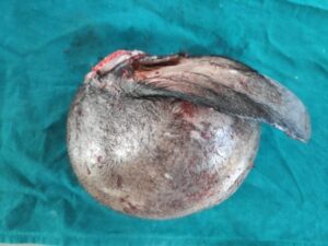
Fig 2: Excised tumour mass |
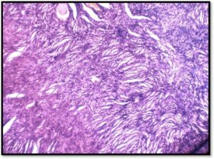
Fig 3: Fibroma showing collagen rich fusiform to spindle shaped hyperchromatic fibroblasts arranged as interlacing bundles and whorls. H&E x200. |