

Surgical Management of Melanoma in a Crossbred Cow
- Vigneswari1*, P. Sankar2, A. Kumerasan2, S. Kathirvel2, K. Jayakumar2 and S. Vigneshwaran2
1Assistant Professor,
Department of Veterinary Surgery and Radiology
Rajiv Gandhi Institute of Veterinary Education and Research (RIVER),
Kurumbapet, Puducherry, India.
2 Department of Veterinary Surgery and Radiology
Veterinary College and Research Institute, Namakkal – 637 002 (Tamil Nadu)
Tamil Nadu Veterinary and Animal Sciences University (TANUVAS), India.
1*Corresponding author
E mail ID: vigneswari.n0@gmail.com
Abstract
A pleuriparous Jersey crossbred cow was presented to the Veterinary Clinical Complex, Veterinary College and Research Institute, Namakkal with the history of swelling at the dorsal aspect of brisket region for past one year. Clinical examination the swelling was hard in consistency. All physiological parameters and haemato-biochemical parameters were within normal range. The mass was tentatively diagnosed as tumour. The tumour was surgically excised under xylazine sedation with local infiltration analgesia. Surgical wound was closed routinely and histopathological examination confirmed the tumour as melanoma. Antibiotic and analgesics were administered postoperatively and daily cutaneous wound dressing was performed. On 10th post-operative day sutures were removed and animal made an uneventful recovery.
Key words: Melanoma; Jersey cross bred cow; brisket region.
Introduction
Melanoma is a benign tumour of melanoblasts, the melanin containing cells, present in the basal layer of the epidermis and mostly seen in dark colored cattle, buffaloes, sheep and goats (Tyagi and Jit Singh, 1996). This tumour is rare in cattle and usually account for 5% to 6% of all tumours in this species. (Patil et al. 2013). Melanomas in cattle occur on head, neck, leg and trunk (Susan and Ducharme, 2017). Tumours are either present at birth or develop within the first 2 years. Melanoma is recognized clinically by areas of gray to black pigmentation within a solid fleshly raised mass. Most melanin pigment tumours are benign in cattle; however, in some cases malignancy may occur. It occurs within the dermis, the subcutis, or both.
Materials and methods
A pleuriparous Jersey crossbred cow was presented with the history of swelling at the dorsal aspect of brisket region for past one year. Clinical examination the swelling was hard in consistency (Figure 1). Physiological and haemato-biochemical parameters were within normal range. The mass was tentatively diagnosed as tumour. The tumour was surgically excised under xylazine sedation with local infiltration analgesia (Figure 2). The tumour mass was sent for histopathological examination and confirmed the tumour as melanoma showing brownish deposits of melanin surrounded by numerous fibroblast and collagen (Figure 3). Surgical wound was closed routinely. Antibiotic and analgesics were administered postoperatively. On 10th post-operative day sutures were removed after complete healing of surgical wound.
Results and Discussion
Melanomas generally originate from neuroectodermal melanoblasts, which migrate at the beginning of the development period into the epidermal- dermal junction of the skin, follicles, and dermis (Susan and Ducharme, 2017). In the present case, it originated from the skin of the brisket region. It is claimed that the majority of melanomas are caused by secondary mutations due to ultra-violet radiation of the types UVA (320-400nm) and UVB (290-320nm) (Slominski, 2001). According to the predominant cell type, melanomas are classified as epithelioid, spindle cell melanomas, mixed and whorled or dendritic melanomas (Gross, 2005), while mixed cell type was observed in the present case. According to Susan and Ducharme 2017, melanomas in cattle are almost always benign. The fact that bovine melanomas can grow for long periods and become massive without killing the animal indicates that most of them are benign. This paper discuss about melanoma in the brisket region of jersey cross bred cow and its surgical management.
References
Cotchin, B. (1960). Tumors of farm animals. Vet. Rec., 72: 816-822.
Gross, T.L. (2005) Melanocytic tumors. In: Gross, T.L. et al. Skin Diseases of the Dog and Cat. Clinical and histopathologic diagnosis. 2nd Edn. Iowa, Blackwell, Pp 813-836.
Patil, A. S., Jadhav Sangeeta and Nagaraj, B.N. (2013). Surgical management of melanoma in a Khillar bullock. Intas Polivet., 14(2): 466 – 468.
Slominski, A. (2001). Malignant melanoma: an update. Arch. Pathol. Lab. Med., 125:1295-1306.
Susan, L. F, and N.G. Ducharme. (2017). Farm Animal Surgery, Second Edition, p. 114.
Tyagi, R.P.S. and Jit Singh. (1996). Digestive system. In Ruminant Surgery, CSB Publishers and distributors, p. 186.
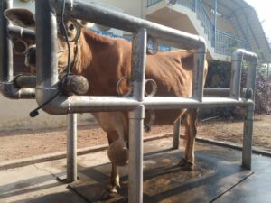
Fig 1: Swelling at the dorsal aspect of brisket region |
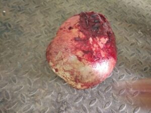
Fig 2: After complete removal of tumour mass |
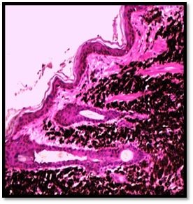
Fig 3: Melanoma showing brownish deposits of melanin surrounded by numerous fibroblast and collagen. Masson’s trichrome x 200
|