

SURGICAL MANAGEMENT OF PERVIOUS URACHUS IN A BUFFALO CALF – A CASE REPORT
- Kumerasan2, M. Vigneswari1*, S. Kathirvel2, P. Sankar2, K. Jayakumar2 and
- Vigneshwaran2
1Assistant Professor,
Department of Veterinary Surgery and Radiology
Rajiv Gandhi Institute of Veterinary Education and Research (RIVER),
Kurumbapet, Puducherry, India.
2 Department of Veterinary Surgery and Radiology
Veterinary College and Research Institute, Namakkal – 637 002 (Tamil Nadu)
Tamil Nadu Veterinary and Animal Sciences University (TANUVAS), India.
*Corresponding author
E mail ID: vigneswari.n0@gmail.com
Abstract
One month old female buffalo calf presented to Veterinary clinical complex, Veterinary College and Research Institute, Namakkal with the history of dribbling of urine from both vulva and umbilicus was diagnosed with pervious urachus a congenital anomaly and it was corrected surgically under general anaesthesia. Normal urination from vulva was found in buffalo calf immediately after surgery and showed no postoperative complications.
Keywords: Pervious Urachus, Buffalo calf
Introduction
During prenatal life, urinary bladder communicates with allantois through a structure known as urachus which becomes atrophied and its lumen gets cicatrized after parturition (Laverty and Salisbury, 2002).Partial or complete failure of lumen obliteration of this structure results in different anomalies like pervious urachus where complete length of urachal lumen fails to obliterate (Langan et al., 2001). The most common congenital condition of the urinary bladder is the pervious urachus. This condition is more commonly observed in the foals (Mc Gavin et al., 2001), cow calves (Dilipkumar and Dhage, 2010) and rare in the buffalo calves (Mouli, 1988 and Sharma and Singh, 2004). In such calves, urine dribbles from the umbilicus and area around it remains wet. Animals with this condition show dribbling of urine from umbilicus and the area surrounding it remains wet (Anjaneya et al., 2016). Usually pervious urachus is accompanied by omphalitis, omphalophlebitis, urachitis and uroperitoneum. The present paper describes the diagnosis and surgical management of pervious urachus in a buffalo calf.
Materials and Methods
One month old female buffalo calf presented to Veterinary clinical complex, Veterinary College and Research Institute, Namakkal with the history of dribbling of urine from both vulva and umbilicus. No signs of omphalitis or any systemic infections were noticed. On clinical examination, umbilical cord thickening with dribbling of urine was noticed (Fig 1). The local area around the umbilicus was inflamed, edematous and pain evinced by the calf on palpation. Caudal to the umbilicus, a hard card like structure could be palpated. In calf, the structures of vulvo-vagina like clitoris, external urethral orifices etc. were normal. Based on the findings of clinical examination, the condition was diagnosed as pervious urachus and it was decided to perform surgical correction. Physiological, haematological and biochemical parameters were within the normal ranges.
Results and Discussion
The calves were prepared for aseptic surgery and premedicated with dexmedetomidine at the dose rate of 1 μg/kg body weight and butorphanol at the dose rate of 0.02mg/kg body weight intravenously and induction and maintenance with ketamine hydrochloride at the dose rate of 2mg/kg body weight. Elliptical incision of skin was given around the umbilicus. After separation of fascia, the urachus and blood vessels were identified through blunt dissection. The urachus was traced up to the bladder and was ligated as close to the bladder as possible (Fig 2) and then the same was resected (Fig 3). The abdominal rent was closed by standard surgical procedure (Fig 4). Post surgery calf was given Streptopencillin @ 100mg/10Kg Bwt I/M O.D for 5 days and Melonex @ 0.2 mg/Kg Bwt I/M O.D was given for 3 days. The animals passed urine from vulva immediate postoperatively and the animal showed uneventful recovery.
Pervious urachus was reported as a common anomaly in domestic foals (Mc Gavin et al., 2001), cow calves (Dilipkumar and Dhage, 2010) and as a rare finding in buffalo calves by (Mouli, 1988 and Sharma and Sing, 2004). Etiological factors responsible for this condition are failure of urachal involution, neonatal omphalitis, umbilical abscess and congenital urethral obstruction (Mc Gavin et al., 2001). In the present study no systemic infections were noticed which could be attributed to their early presentation. Contrary to this, Anjaneya et al., (2016) observed uroperitoneum, cystitis, umbilical cord infection and urinary obstruction in a cow calf suffering from this condition. If the urachus remains open for longer period it may act as a source of ascending infections to the bladder (Langan et al., 2001) which was not found in any of the cases under present study.
Urethra was considered to be patent as the animal was voiding urine through urethral opening at vulva besides urachal opening at umbilicus. Prognosis of pervious urachus condition is grave when the urethra is permanently occluded (O`Connor, 1980). In the present study authors ascertained the condition of urethra in the calf before the initiation of treatment. Urachus was ligated close to the bladder and excised as suggested by Oehme and Prier, (1974) to treat the animals suffering from this condition.
Treatment procedures like application of blister around the orifice, needle point firing, ligation of urachus, inserting a needle suture were advised by O`Connor, (1980) to treat pervious urachus condition where as Sharma and Singh, (2004) suggested application of 90% phenol dipped cotton swab over the urachus as a conservative method of treatment. As the farmers who own these animals wanted a permanent remedy and as there were no complications, conservative therapy could not be even thought off and surgery was resorted to. Earlier diagnosis and surgical resection of patent urachus in a buffalo calf ensured a good recovery. Pervious urachus is a congenital anomaly reported in young calves characterized by voiding of urine both through the vulval and umbilical openings. This should be treated surgically as early as possible by resecting the urachus to avoid ascending infections to bladder and peritoneum.
References
Anjaneya, S.N., A. Bhimappa, R.R. Reddy, B.V. Shivaprakash and P. Udagatti. 2016. Pervious urachus in calves and its surgical management. Int. J of Science. Env and Tech., 5: 730 – 732.
Dilipkumar., D. And G.P. Dhage. 2010. Surgical treatment of pervious urachus in calves. Journal of Vet. Pract., 11: 39.
Langan, J., E. Ramsay, J. Schumacher, T. Chism, and S. Adair. 2001. Diagnosis and management of a patent urachus in a white rhinoceros calf (Ceratotherium simum simum). Journal of Zoo and Wildlife Medicine, 32: 118–122.
Laverty, P. H and S. K. Salisbury. 2002. Surgical management of true patent urachus in a cat. J. Small. Anim. Pract., 43: 227-229.
McGavin, M.D., W.W. Carlton and J.F. Zachary. 2001. The urinary system. In:Thomson’s Special Veterinary Pathology (3rd Edn). P 271-272.
Mouli, S.P. 1998. Surgical therapy of pervious urachus at the umbilicus in a neonatal graded buffalo bull-calf. Indian Vet. J., 65:75.
O’Connor, J.J. 1980. Dollars Veterinary Surgery, general,operative and regional, 4th Edtn. CBS Publishers, New Delhi. Page no 738-739.
Oehme, F.W. and J.E. Prier. 1974. Text book of Large Animal Surgery. Williams & ilkins, Baltimore, U.S.A, pp.447-448.
Sharma, S.N. and J. Singh. 2004. The urinary system. In: Ruminat Surgery, R.P.S. Tygi and Jit Singh (Eds). C.B.S. Publishers and Distributrs, New Delhi. P 272.
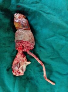
Figure 3: Resected urachus and umbilical vessels |
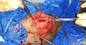
Figure 4: Abdominal rent closure |
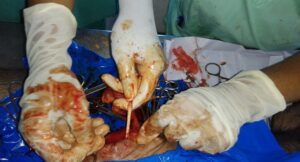
Figure 2: Placement of ligature to the urachus |
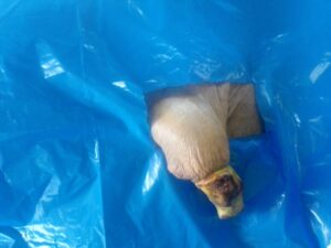
Figure 1: Umbilical cord thickening |