

Surgical Management of Choke in a Gir Cow at Field Condition
Dr. M. Vigneswari
Field Veterinarian,
Karaikal (district), Pondicherry (state) – 609605
Abstract
A four years old Gir cow was presented with a history of excessive salivation, restlessness, inability to swallow feed or water, severe bloat and palpable swelling at the left lateral cervical part of the oesophagus. It was diagnosed as a cervical oesophageal obstruction (choke) by passing probang. Oesophagotomy was performed and foreign body (Toddy palm fruit) was successfully removed at the field condition with limited facilities.
Keywords: Choke, Oesophageal obstruction, Toddy palm fruit, Gir Cow.
Introduction
In cattle, acute and complete oesophageal obstruction is an emergency condition because it prohibits the eructation of ruminal gases leading to severe free gas bloat which may be life threatening if not relieved in time (Prakash et al, 2014). The common sites of obstruction in bovines include pharynx, cervical oesophagus, thoracic inlet, base of heart and cardiac (Thyagi and Singh, 1999). Diagnosis of choke depends on history of eating habits and clinical signs as bloat, tenesmus, retching and salivation. External palpation may be used to confirm those located in cervical oesophagus. The additional diagnostic tools that may help to determine the location of obstruction include oral explorations, passing probangs or stomach tubes, oesophageal endoscopy and radiography of oesophagus. The primary indication for oesophageal surgery in large animals is to relieve oesophageal obstructions (choke) which have not responded to conservative treatment (Meagher and Mayhew, 1978). In the present case, successful surgical management of choke at field condition with limited facility is discussed.
————————————————————————————————————
* Corresponding author:
Field Veterinarian, Karaikal (district), Pondicherry (state),
Email: vigneswari.n0@gmail.com
Phone No: 7550282548
Case history and Observation
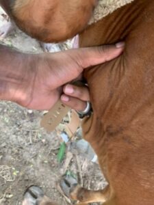
A four years old Gir cow was presented in the field condition in the area of Karaikal district, with the history of excessive salivation, restlessness, inability to swallow feed or water, severe bloat and palpable swelling at the left lateral cervical part of the oesophagus. On palpation a rigid hard mass (Fig.1) was observed on left ventro-lateral aspect of mid cervical region. Clinical examination revealed that the rectal temperature was within normal range, respiratory rate was 64 breaths /min and heart beat was 114 beats /min. Attempts to relive choke by pushing into rumen using probang under Xylazine sedation were unsuccessful. Based on history and severe respiratory distress of animal, it was decided to perform cervical oesophagotomy.
Treatment and Discussion
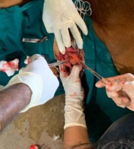
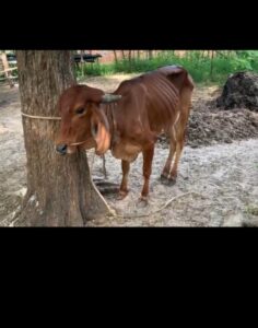
Trocar and canula was applied on left paralumbar fossa after preparing aseptic site to relieve bloat till completion of surgery. Animal was restrained in right lateral recumbency and surgical site was prepared aseptically under xylaxine sedation at the dose rate of 0.1mg/kg body weight intramuscularly. Local analgesia was achieved by 2% lignocaine hydrochloride which was infiltrated around the swelling. A10 cm linear incision was made on the left side over the mass and along the dorsal aspect of the jugular furrow. Care was taken not to puncture the jugular vein and artery. The oesophagus was approached between the sterno-cephalicus muscle and trachea. The operative site was packed with sterile gauge to avoid any contamination by the fluid in the oesophagus. A 5 cm longitudinal incision was made through the lateral wall of the esophagus and the foreign body (Toddy palm fruit – nonku) was removed (Fig. 2). The mucosal layer was sutured with simple interrupted sutures utilizing chromic catgut No. 1 as intraluminal sutures. The submucosa and muscularis were closed in one layer using a simple continuous suture pattern with No. 1 chromic catgut. The muscles and skin were closed in routine manner. Postoperatively, animal was administered with fluid therapy twice daily for 5days, inj. streptopenicillin 5gm and inj. meloxicam 10 ml intramuscularly for five and three days, respectively. The cutaneous sutures were removed on 10th postoperative day (Fig 3).
Bovines are frequently affected by oesophageal obstruction than other animals and this is attributable to their greedy nature and peculiar indiscriminate feeding habits. Attempts to push the object with a probang were not successful, perhaps due to a change in position of kernel at the site. Oesophageal obstruction due to palm kernel (Hari Krishna et al, 2011), mango (Veena et al, 2000), tarpaulin cloth (Sreenu and Sureshkumar, 2001) has been corrected surgically without any complication. In the present case, toddy palm fruit was retrieved from the oesophagus. It may be due to the availability of palm fruit in that area and swallowed by the animal sent for grazing. Ruben, (1997) reported the risk of postoperative complications associated with an oesophagotomy as incisional dehiscence and fistula formation but in present case, no such raw complications were seen and animal made an uneventful recovery in the field condition.
Conflict of interests
The authors declared no potential conflicts of interest with respect to the clinical work, authorship, and/or publication of this article.
References
Hari Krishna, N.V.V. Makkena, S and Bose, V.S.C. (2011). An unusual case of oesophageal obstruction in a female buffalo. Buffalo Bulletin, 30:4-5.
Meagher, D. M. and Mayhew, I. G. 1978. The surgical treatment of upper oesophageal obstruction in the bovine. Can. vet. J., 19; 128-132.
Ruben., J. M. (1997). Surgical removal of a foreign body from the bovine oesophagus. Vet Rec. 100:220.
Sreenu M, Sureshkumar RV (2001). Obstruction of oesophagus by tarpaulin cloth in a buffalo calf. Indian Vet. J., 78: 243-244.
Tyagi, R.P.S. and Singh, J. (1999). Ruminant Surgery. Ist Edn. CBS Publishers and Distributers, New Delhi, India. Pp- 192.
Veena P, Ravikumar A, Ramakrishna O (2000). Oesophageal obstruction by a mango in a heifer. Indian Vet. J., 77: 794.
|
|
|