


SURGICAL MANAGEMENT OF DYSTOCIA IN ELEPHANT (Elephas Maximus)
Abstract:
This case was presented with history of showing signs of parturition for 2days in 13yrs aged female elephant from Champua Range of Keonjhar forest division. Later on it was observed, two hind limbs of a full-term dead calf advanced into and lodged outside the vagina. The diagnosis was dystocia. And the decision was taken to tranquilize the elephant and attempt to pull out the dead edeamatous foetus by traction. At the end of the surgical procedure and uneventful recovery was observed.
Key word: Dystocia, Edematous foetus, Elephas maximus
Introduction: The elephant is the largest land mammal with the longest gestation period of 20-22 months. Unique anatomical features of the female elephant include:
- Long reproductive tract over 3 m from vestibule to ovary,
- ventrally directed opening of the vulva which is located between the hind limbs,
- permanent hymeneal membrane in nulliparous females which is not ruptured during natural mating,
- Small vaginal opening with two blind pouches of similar size on either side of it.
In part, arising from these anatomical features, dystocia and stillbirths are major issues in elephants in captivity.
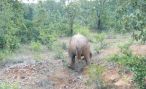
Case History: A female elephant aged about 13yrs from Champua range of Keonjhar forest division, Odisha was reported having difficulty in labor (dystocia). The elephant showed signs of parturition for 2 days and later found two hindlimb of a full term dead calf advanced into and lodged outside the vagina. Edematous swelling of the whole foetal body and bad odour was observed.
|
|
 |

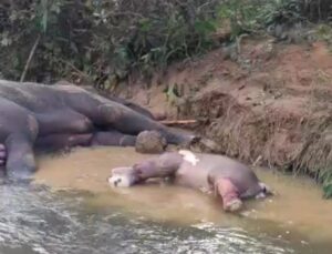
Treatment: Prior to beginning of the treatment, the elephant was tranquilized by darting Inj. Xylazine HCl (300mg) and Inj. Ketamine HCl (200mg) intramuscularly. Maintenance was done by administration of top up mixture of Inj. Xylazine and Inj. Ketamine intravenously throughout the procedure. Continuous IV fluid was given and analgesics was given as soon as the elephant was sedated. The foetus was in posterior presentation. Edematous swelling of the whole foetal body and bad odour was observed. Manual traction was applied at first via a rope tied on the hind limb. After several attempt, it was proved in sufficient to achieve delivery by traction & planed for episiotomy. Local anaesthesia using 2% lidocaine hydrochloride was infiltrated along the proposed incision line. A 32 cm long incision was made in the perineum. Traction was applied through ropes that were attached to the hind limbs but still it was difficult to pull out the foetus as calf was already in edematous condition. At this point, the incision was extended by approximately 10cm. This time the calf was successfully extracted by pulling on either the two ropes on the hind limbs. Considerable fibrous tissue and clotted blood were observed together with a laceration in the ventral aspect of the cervix. Povidone iodine was used to flush the debris and fibrous tissue from the uterus. The episiotomy incision was then sutured in two layers of simple continuous sutures using chromic catgut no.2 to close perineal muscle and subcutis. The skin was sutured in routine manner using trulon no.2-0.
 |
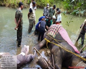 |
| EXTRACTION OF FETUS AND CLOSURE OF INCISIONAL WOUND | |
Then sulfadimethylpyrimidine-trimethoprim boluses were introduced into the uterine lumen. Antiseptic ointment was applied on wound externally. Systemic antibiotic was given intramuscularly and the sedation was then reversed by intravenous administration of 150mg yohimbine hydrochloride at the end of the surgical procedure. Post-operative care was taken till healing of the wound.
Conclusion:
Dystocia has been reported in both Asian and African elephants but appears to be more common in the Asian species. But the underlying causes of prolonged foetal retention in the elephant are unknown. In the current case, oxytocin was not used to stimulate uterine contractions because the history of prolonged fetal retention made it more likely that there would be insufficient amniotic fluid to lubricate fetal passage and therefore, the risk of oxytocin-induced uterine rupture was considered to be higher than normal. Furthermore, studies related to initial causes, possible treatment of, failure of parturition to progress, birth management protocols and active training of veterinary teams in how to manage dystocia in elephants would help to improve the outcome in many cases.