

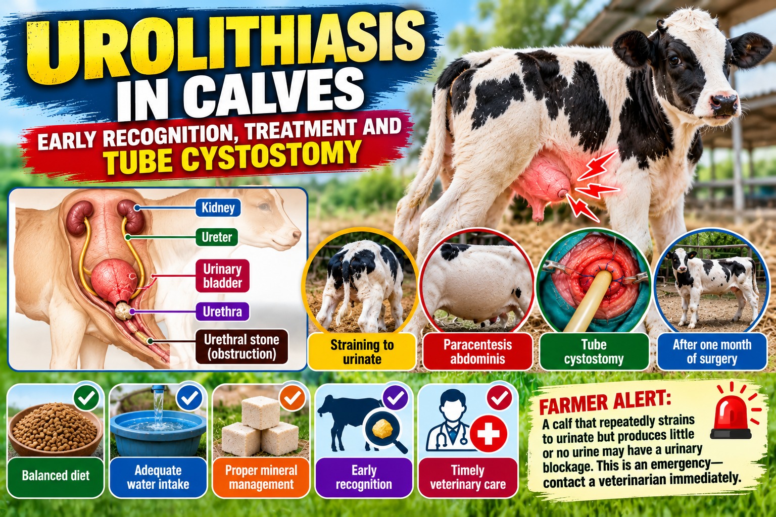
UROLITHIASIS IN CALVES: EARLY RECOGNITION, TREATMENT AND TUBE CYSTOSTOMY
Dr. Supreetkumar Sagar, M.V.Sc., NET, PGDAW1 and Dr. Sanjeev S. Pitlawar, M.V.Sc., PhD, PGDAW2
1: Veterinary Surgeon (Gold medalist), ALUMNUS,College of Veterinary and Animal Sciences, Udgir, Maharashtra Animal & Fishery Sciences University, Nagpur
2: Associate Professor and Sectional Head, Department of Veterinary Surgery and Radiology, College of Veterinary and Animal Sciences, Udgir, Maharashtra Animal & Fishery Sciences University, Nagpur
| FARMER ALERT: A calf that repeatedly strains to urinate but produces little or no urine may have a urinary blockage. This is an emergency—contact a veterinarian immediately. |
Introduction
Urolithiasis, commonly called urinary stone disease, is an important condition in young male calves, particularly those receiving concentrate-rich diets. Mineral deposits may form stones (uroliths) within the urinary tract. When a stone obstructs the urethra, urine cannot pass normally and the bladder may become severely distended or rupture.
The condition is particularly important in castrated male calves because the urethra is relatively narrow and the risk of obstruction may be increased. Early recognition is essential because a complete obstruction can rapidly become life-threatening.
What Causes Urinary Stones?
Urolith formation is influenced by several interacting factors rather than a single cause. These include the mineral composition of the diet, urine concentration, water intake, urinary pH, growth rate and management.
- High-concentrate diets with inadequate roughage
- Imbalance of dietary calcium and phosphorus
- Excessive intake of certain mineral sources
- Low water intake or restricted access to clean water
- Rapid changes in diet
- Poor-quality or improperly balanced calf starter/concentrate
- Conditions that produce concentrated urine
- Insufficient dietary management of urinary pH where appropriate
- Individual susceptibility and previous urinary problems
Incidence and High-Risk Calves
The occurrence of urolithiasis varies widely between farms and depends on feeding, mineral balance, water availability, management and animal factors. Therefore, a single incidence percentage should not be assumed for every farm. A rising number of cases should prompt a review of the farm’s feeding and mineral programme.
- Male calves, especially castrated males
- Calves receiving large amounts of concentrate
- Calves with limited access to fresh drinking water
- Animals exposed to sudden dietary changes
- Groups with a previous history of urinary blockage
Early Clinical Signs
Farmers should watch young male calves closely. The earliest signs may be mistaken for constipation or abdominal pain.
- Repeated straining to urinate
- Frequent attempts to urinate with little or no urine
- Tail switching or repeated lifting of the tail
- Restlessness and kicking at the abdomen
- Grinding of teeth due to pain
- Vocalization or unusual posture
- Dribbling of urine
- Swelling around the preputial region
- Reduced appetite and depression
Advanced Signs and Complications
With complete obstruction, the bladder can become markedly distended. Continued accumulation of urine may result in rupture of the bladder or, depending on the site of obstruction, leakage of urine into surrounding tissues. Such calves may become depressed, dehydrated and weak and require emergency veterinary intervention.
Diagnosis
Diagnosis is based on history and clinical examination. The veterinarian may assess the urinary bladder and urethra by palpation and ultrasonography and may perform additional diagnostic tests where required. Blood examination can help assess dehydration and disturbances associated with urinary obstruction. The location and severity of obstruction determine the most appropriate treatment.
Treatment: Act Early
Urolithiasis with urinary obstruction is a veterinary emergency. Treatment aims to restore urine flow, relieve pressure on the urinary tract, correct dehydration and electrolyte disturbances, and prevent recurrence.
Immediate Veterinary Management
- Assess the degree and location of urinary obstruction.
- Relieve the obstruction where medically and surgically appropriate.
- Provide intravenous or other appropriate fluid therapy when indicated.
- Correct dehydration and electrolyte/acid–base abnormalities.
- Provide appropriate analgesic and anti-inflammatory support.
- Monitor urine production and the calf’s general condition closely.
Tube Cystostomy: A Life-Saving Surgical Option
When urethral obstruction cannot be safely or successfully relieved, or when the urinary bladder is severely affected, a veterinarian may perform tube cystostomy. In this procedure, a catheter/tube is surgically placed into the urinary bladder through the abdominal wall to provide an alternative route for urine drainage.
Tube cystostomy bypasses the obstructed urethra and allows urine to leave the bladder while the urethral obstruction is managed. It is particularly useful in selected calves with persistent obstruction and is generally preferred to repeatedly forcing instruments through a damaged urethra.
Basic Principles of Tube Cystostomy
- The calf is appropriately restrained and prepared for surgery using strict aseptic technique.
- The urinary bladder is identified and accessed by the veterinarian at an appropriate site.
- A suitable urinary catheter/tube is securely placed into the bladder.
- The tube is fixed to prevent leakage or accidental removal.
- Urine is diverted through the tube while the primary obstruction is managed.
- The surgical site and tube are monitored regularly for infection, leakage and displacement.
Aftercare Following Tube Cystostomy
- Keep the surgical site clean and dry.
- Prevent the calf from damaging or pulling the tube.
- Monitor urine flow through the tube.
- Observe for fever, swelling, discharge, abdominal pain or reduced urine flow.
- Provide prescribed analgesics, antibiotics or other medicines only as directed by the veterinarian.
- Continue monitoring normal urination as the urethra recovers.
- Do not remove or clamp the tube unless instructed by the veterinarian.
| WARNING | WHY IT MATTERS |
| Repeated straining with no urine | May indicate complete obstruction and requires immediate attention. |
| Abdominal enlargement/pain | May indicate severe bladder distension or urinary leakage. |
| Sudden depression or weakness | May occur with advanced obstruction and metabolic complications. |
| No urine through a cystostomy tube | May indicate blockage, kinking or displacement of the tube. |
Prevention: The Most Economical Approach
Prevention depends mainly on correct feeding, mineral balance, adequate water intake and close observation of young male calves.
- Provide unlimited access to clean, fresh drinking water.
- Feed a balanced calf ration appropriate for age and growth rate.
- Avoid excessive concentrate feeding and sudden dietary changes.
- Maintain an appropriate balance of dietary calcium and phosphorus.
- Do not provide mineral supplements indiscriminately; use a properly formulated ration.
- Ensure adequate roughage according to the calf’s age and feeding programme.
- Maintain clean feed and water troughs.
- Monitor groups with a previous history of urolithiasis more closely.
- Seek veterinary/nutritional advice when multiple cases occur.
Farm Management Checklist
| Area | What to Check | Action |
| Water | Fresh, clean water available throughout the day | Clean troughs and ensure continuous access |
| Feed | Concentrate level and roughage supply | Use a balanced ration; avoid sudden changes |
| Minerals | Calcium, phosphorus and other mineral sources | Review ration with a veterinarian/nutritionist |
| Calves | Straining, dribbling or reduced urination | Call veterinarian immediately |
| Previous cases | History of obstruction in the group | Increase monitoring and review feeding programme |
| Surgical cases | Tube, urine flow and incision | Follow veterinary aftercare instructions |
| REMEMBER: DO NOT WAIT FOR COMPLETE URINARY BLOCKAGE. Early veterinary examination can prevent bladder damage, rupture and death. Tube cystostomy is a surgical procedure performed by a veterinarian when urinary diversion is required. |
||
Common Mistakes to Avoid
- Do not assume repeated straining is constipation.
- Do not wait for the calf to stop eating before seeking help.
- Do not forcefully attempt to rupture or push a suspected stone without veterinary assessment.
- Do not give large volumes of fluid to an obstructed calf without veterinary guidance.
- Do not use unprescribed medicines or human urinary medicines.
- Do not ignore reduced urine output after surgery.
- Do not remove a cystostomy tube without veteran
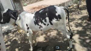
Young male calf showing typical straining posture during urinary obstruction. |
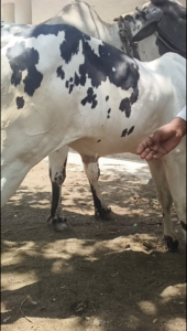
Paracentesis Abdominis |
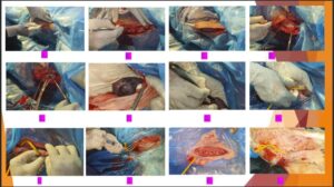
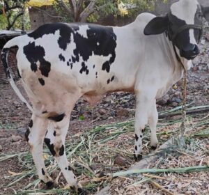
Tube cystostomy procedure After One month of surgery
Conclusion
Urolithiasis is a preventable but potentially fatal problem in young male calves. The key to successful management is early recognition of abnormal urination and immediate veterinary intervention. Balanced feeding, adequate water intake, appropriate mineral management and careful monitoring can reduce the risk of urinary stone formation. When obstruction persists, tube cystostomy can provide an effective urinary diversion route and give the calf an opportunity to recover while the underlying urethral problem is managed.
“Early detection saves the bladder—and can save the calf.”
References
- Radostits, O.M., Gay, C.C., Hinchcliff, K.W. and Constable, P.D. Veterinary Medicine: A Textbook of the Diseases of Cattle, Horses, Sheep, Pigs and Goats.
- Smith, B.P. Large Animal Internal Medicine. Elsevier.
- Fubini, S.L. and Ducharme, N.G. Farm Animal Surgery. Elsevier.
- Constable, P.D., Hinchcliff, K.W., Done, S.H. and Grünberg, W. Veterinary Medicine: Diseases of Cattle, Horses, Sheep, Pigs and Goats.